<< Chapter < Page Chapter >> Page >
Illustration shows cells, shaped like slices of pie, arranged in a circle. The hub of the circle is empty. Three of these circles of cells cluster together.
Simple cuboidal epithelial cells line tubules in the mammalian kidney, where they are involved in filtering the blood.

Columnar epithelia

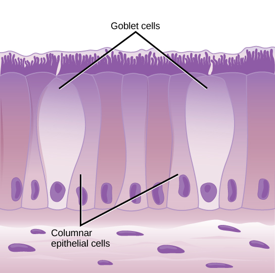
Columnar epithelial cells are taller than they are wide: they resemble a stack of columns in an epithelial layer, and are most commonly found in a single-layer arrangement. The nuclei of columnar epithelial cells in the digestive tract appear to be lined up at the base of the cells, as illustrated in [link] . These cells absorb material from the lumen of the digestive tract and prepare it for entry into the body through the circulatory and lymphatic systems.

Illustration shows tall, columnar cells arranged side-by-side. Each cell has a nucleus located near the bottom, and cilia extending from the top. Two oval goblet cells are interspersed among the columnar epithelial cells. The goblet cells, which are shorter than the columnar cells, are in direct contact with the intestinal lumen. Beneath the columnar cells is a layer of horizontal cells.
Simple columnar epithelial cells absorb material from the digestive tract. Goblet cells secret mucous into the digestive tract lumen.

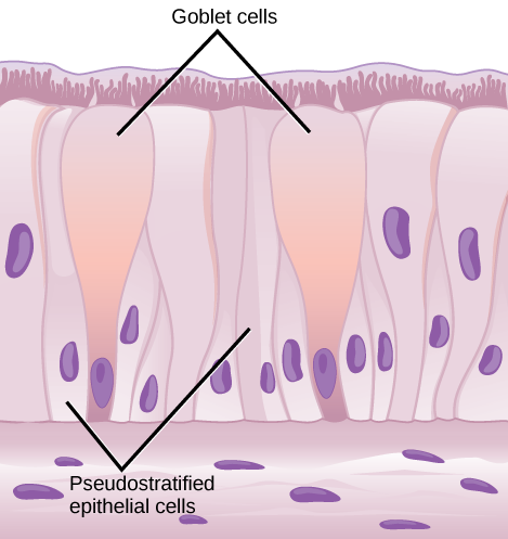
Columnar epithelial cells lining the respiratory tract appear to be stratified. However, each cell is attached to the base membrane of the tissue and, therefore, they are simple tissues. The nuclei are arranged at different levels in the layer of cells, making it appear as though there is more than one layer, as seen in [link] . This is called pseudostratified    , columnar epithelia. This cellular covering has cilia at the apical, or free, surface of the cells. The cilia enhance the movement of mucous and trapped particles out of the respiratory tract, helping to protect the system from invasive microorganisms and harmful material that has been breathed into the body. Goblet cells are interspersed in some tissues (such as the lining of the trachea). The goblet cells contain mucous that traps irritants, which in the case of the trachea keep these irritants from getting into the lungs.

Illustration shows columnar cells arranged side-by-side. The cells are wide at the top, and thin at the bottom. Shorter columnar cells are interspersed between the lower, thin part of the tall columnar cells. Some of these cells extend to the surface of the epithelium, but they are very thin at the top. The nuclei of the tall columnar cells are located near the top, and the nuclei of the shorter columnar cells are located near the bottom, giving the appearance of two layers of cells. Cilia extend from the top of the tall columnar cells. Oval goblet cells are interspersed among the columnar epithelial cells. Beneath the columnar cells is a layer of horizontal cells.
Pseudostratified columnar epithelia line the respiratory tract. They exist in one layer, but the arrangement of nuclei at different levels makes it appear that there is more than one layer. Goblet cells interspersed between the columnar epithelial cells secrete mucous into the respiratory tract.

Transitional epithelia

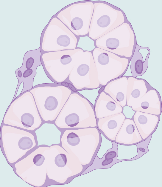
Transitional or uroepithelial cells appear only in the urinary system, primarily in the bladder and ureter. These cells are arranged in a stratified layer, but they have the capability of appearing to pile up on top of each other in a relaxed, empty bladder, as illustrated in [link] . As the urinary bladder fills, the epithelial layer unfolds and expands to hold the volume of urine introduced into it. As the bladder fills, it expands and the lining becomes thinner. In other words, the tissue transitions from thick to thin.

Art connection

Illustration shows tall, diamond-shaped cells layered one on top of the other.
Transitional epithelia of the urinary bladder undergo changes in thickness depending on how full the bladder is.

Which of the following statements about types of epithelial cells is false?

  1. Simple columnar epithelial cells line the tissue of the lung.
  2. Simple cuboidal epithelial cells are involved in the filtering of blood in the kidney.
  3. Pseudostratisfied columnar epithilia occur in a single layer, but the arrangement of nuclei makes it appear that more than one layer is present.
  4. Transitional epithelia change in thickness depending on how full the bladder is.

Questions & Answers

Discuss the differences between taste and flavor, including how other sensory inputs contribute to our  perception of flavor.
John Reply
taste refers to your understanding of the flavor . while flavor one The other hand is refers to sort of just a blend things.
Faith
While taste primarily relies on our taste buds, flavor involves a complex interplay between taste and aroma
Kamara
which drugs can we use for ulcers
Ummi Reply
omeprazole
Kamara
what
Renee
what is this
Renee
is a drug
Kamara
of anti-ulcer
Kamara
Omeprazole Cimetidine / Tagament For the complicated once ulcer - kit
Patrick
what is the function of lymphatic system
Nency Reply
Not really sure
Eli
to drain extracellular fluid all over the body.
asegid
The lymphatic system plays several crucial roles in the human body, functioning as a key component of the immune system and contributing to the maintenance of fluid balance. Its main functions include: 1. Immune Response: The lymphatic system produces and transports lymphocytes, which are a type of
asegid
to transport fluids fats proteins and lymphocytes to the blood stream as lymph
Adama
what is anatomy
Oyindarmola Reply
Anatomy is the identification and description of the structures of living things
Kamara
what's the difference between anatomy and physiology
Oyerinde Reply
Anatomy is the study of the structure of the body, while physiology is the study of the function of the body. Anatomy looks at the body's organs and systems, while physiology looks at how those organs and systems work together to keep the body functioning.
AI-Robot
what is enzymes all about?
Mohammed Reply
Enzymes are proteins that help speed up chemical reactions in our bodies. Enzymes are essential for digestion, liver function and much more. Too much or too little of a certain enzyme can cause health problems
Kamara
yes
Prince
how does the stomach protect itself from the damaging effects of HCl
Wulku Reply
little girl okay how does the stomach protect itself from the damaging effect of HCL
Wulku
it is because of the enzyme that the stomach produce that help the stomach from the damaging effect of HCL
Kamara
function of digestive system
Ali Reply
function of digestive
Ali
the diagram of the lungs
Adaeze Reply
what is the normal body temperature
Diya Reply
37 degrees selcius
Xolo
37°c
Stephanie
please why 37 degree selcius normal temperature
Mark
36.5
Simon
37°c
Iyogho
the normal temperature is 37°c or 98.6 °Fahrenheit is important for maintaining the homeostasis in the body the body regular this temperature through the process called thermoregulation which involves brain skin muscle and other organ working together to maintain stable internal temperature
Stephanie
37A c
Wulku
what is anaemia
Diya Reply
anaemia is the decrease in RBC count hemoglobin count and PVC count
Eniola
what is the pH of the vagina
Diya Reply
how does Lysin attack pathogens
Diya
acid
Mary
I information on anatomy position and digestive system and there enzyme
Elisha Reply
anatomy of the female external genitalia
Muhammad Reply
Organ Systems Of The Human Body (Continued) Organ Systems Of The Human Body (Continued)
Theophilus Reply
what's lochia albra
Kizito
Got questions? Join the online conversation and get instant answers!
Jobilize.com Reply

Get Jobilize Job Search Mobile App in your pocket Now!

Get it on Google Play Download on the App Store Now


Source:  OpenStax, Biology. OpenStax CNX. Feb 29, 2016 Download for free at http://cnx.org/content/col11448/1.10
Google Play and the Google Play logo are trademarks of Google Inc.

Notification Switch

Would you like to follow the 'Biology' conversation and receive update notifications?

Ask