<< Chapter < Page Chapter >> Page >
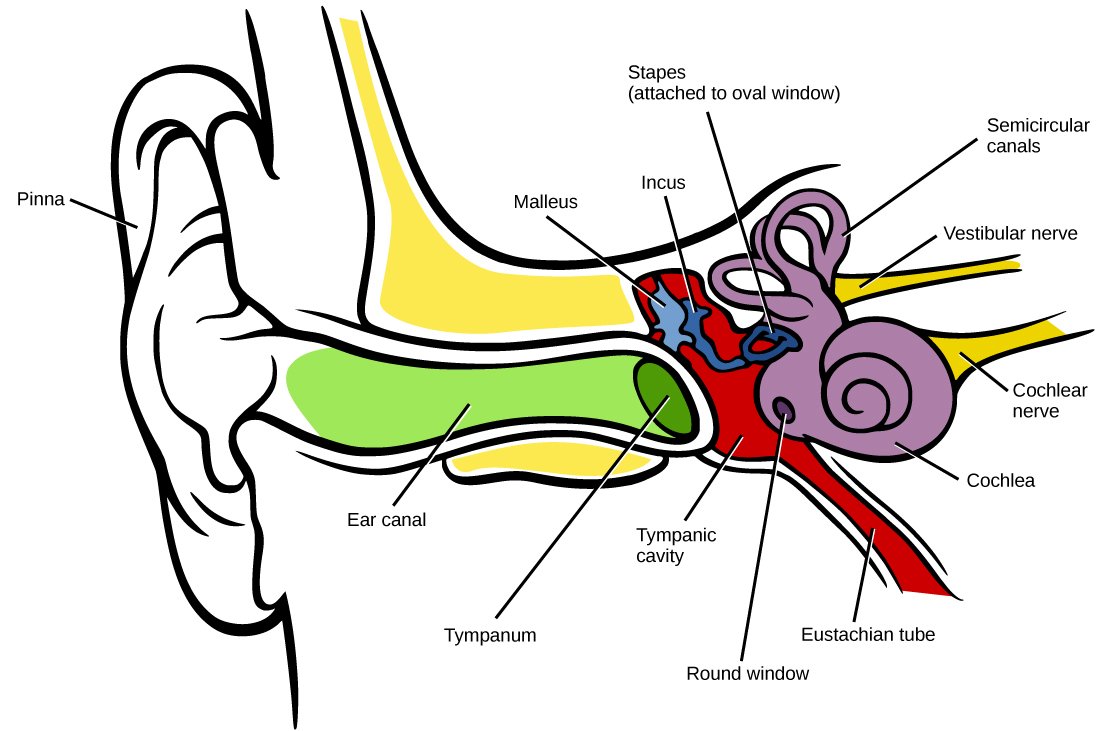
The illustration shows the parts of the human ear. The visible part of the exterior ear is called the pinna. The ear canal extends inward from the pinna to a circular membrane called the tympanum. On the other side of the tympanum is the Eustachian tube. Inside the Eustachian tube the malleus, which touches the inside of the tympanum, is attached to the incus, which is in turn attached to the horseshoe-shaped stapes. The stapes is attached to the round window, a membrane in the snail shell-shaped cochlea. Another window, called the round window, is located in the wide part of the cochlea. Ring-like semicircular canals extend from the cochlea. The cochlear nerve and vestibular nerve both connect to the cochlea.
Sound travels through the outer ear to the middle ear, which is bounded on its exterior by the tympanic membrane. The middle ear contains three bones called ossicles that transfer the sound wave to the oval window, the exterior boundary of the inner ear. The organ of Corti, which is the organ of sound transduction, lies inside the cochlea. (credit: modification of work by Lars Chittka, Axel Brockmann)

Transduction of sound

Vibrating objects, such as vocal cords, create sound waves or pressure waves in the air. When these pressure waves reach the ear, the ear transduces this mechanical stimulus (pressure wave) into a nerve impulse (electrical signal) that the brain perceives as sound. The pressure waves strike the tympanum, causing it to vibrate. The mechanical energy from the moving tympanum transmits the vibrations to the three bones of the middle ear. The stapes transmits the vibrations to a thin diaphragm called the oval window    , which is the outermost structure of the inner ear    . The structures of the inner ear are found in the labyrinth    , a bony, hollow structure that is the most interior portion of the ear. Here, the energy from the sound wave is transferred from the stapes through the flexible oval window and to the fluid of the cochlea. The vibrations of the oval window create pressure waves in the fluid (perilymph) inside the cochlea. The cochlea    is a whorled structure, like the shell of a snail, and it contains receptors for transduction of the mechanical wave into an electrical signal (as illustrated in [link] ). Inside the cochlea, the basilar membrane    is a mechanical analyzer that runs the length of the cochlea, curling toward the cochlea’s center.

The mechanical properties of the basilar membrane change along its length, such that it is thicker, tauter, and narrower at the outside of the whorl (where the cochlea is largest), and thinner, floppier, and broader toward the apex, or center, of the whorl (where the cochlea is smallest). Different regions of the basilar membrane vibrate according to the frequency of the sound wave conducted through the fluid in the cochlea. For these reasons, the fluid-filled cochlea detects different wave frequencies (pitches) at different regions of the membrane. When the sound waves in the cochlear fluid contact the basilar membrane, it flexes back and forth in a wave-like fashion. Above the basilar membrane is the tectorial membrane    .
Art connection

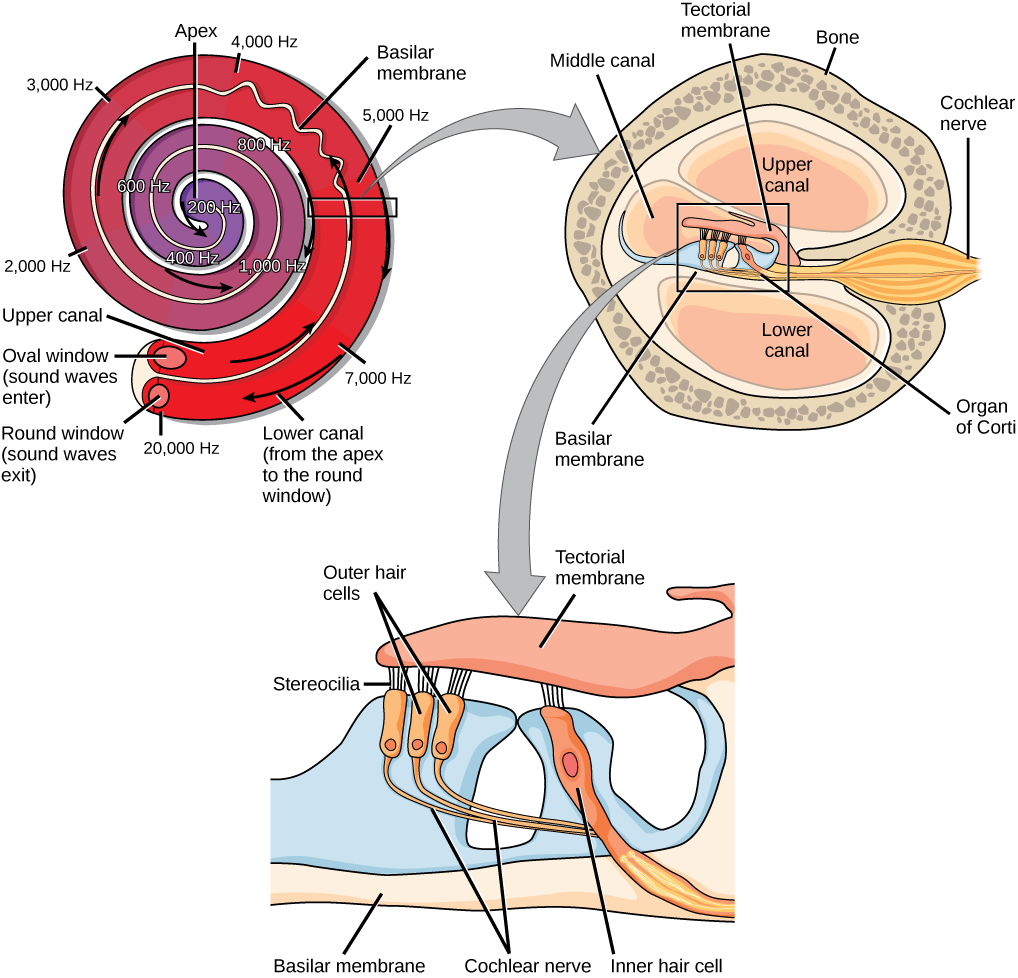
 A series of three illustrations are shown. The top illustration shows a cochlea, which is shaped like a snail shell with two parallel chambers, the upper chamber and the lower chamber, coiling from the outside in. These chambers are separated by a flexible membrane basilar membrane. The oval window covers the inner of these parallel chambers. Sound waves enter here, and travel to the middle, or apex, of the coil. The membrane separating the two chambers gets thinner from the outside in, such that is vibrates at different sound frequencies, about 20,000 hertz on the outside and about 200 hertz on the inside. Sound then travels back out through the lower chamber, and exits through the round window. The middle illustration shows a closer view of a cross-sectional image of the cochlea. A roughly circular shape has a roughly circular bone exterior, with the middle portion of the circle divided into four major areas. Two of these are spaces labeled “upper canal” and “lower canal.” In the middle is the organ of Corti, and extending from the middle out through the outer bone area is the cochlear nerve, which extends from the middle as a thin tube and then bulges into a larger oval shape as it extends through the bone. The bottom illustration is an enlarged image of the organ of Corti. In the view shown, the top section is a flattish pink area called the tectorial membrane. Extending beneath that membrane are three areas with hair-like connectors (stereocilia) that run from the membrane to the outer hair cells. The outer hair cells are shaped like rectangles with rounded corners. From the end of each protrudes a narrow tube: the cochlear nerve. These narrow tubes join to an inner hair cell, which looks similar to the outer hair cells but with its rectangular shape remaining a consistent width instead of narrowing into a nerve. At the bottom of the image, opposite the top tectorial membrane, is a basilar membrane.
In the human ear, sound waves cause the stapes to press against the oval window. Vibrations travel up the fluid-filled interior of the cochlea. The basilar membrane that lines the cochlea gets continuously thinner toward the apex of the cochlea. Different thicknesses of membrane vibrate in response to different frequencies of sound. Sound waves then exit through the round window. In the cross section of the cochlea (top right figure), note that in addition to the upper canal and lower canal, the cochlea also has a middle canal. The organ of Corti (bottom image) is the site of sound transduction. Movement of stereocilia on hair cells results in an action potential that travels along the auditory nerve.

Cochlear implants can restore hearing in people who have a nonfunctional cochlear. The implant consists of a microphone that picks up sound. A speech processor selects sounds in the range of human speech, and a transmitter converts these sounds to electrical impulses, which are then sent to the auditory nerve. Which of the following types of hearing loss would not be restored by a cochlear implant?

  1. Hearing loss resulting from absence or loss of hair cells in the organ of Corti.
  2. Hearing loss resulting from an abnormal auditory nerve.
  3. Hearing loss resulting from fracture of the cochlea.
  4. Hearing loss resulting from damage to bones of the middle ear.

Questions & Answers

it is the relatively stable flow of income
Chidubem Reply
what is circular flow of income
Divine Reply
branches of macroeconomics
SHEDRACK Reply
what is Flexible exchang rate?
poudel Reply
is gdp a reliable measurement of wealth
Atega Reply
introduction to econometrics
Husseini Reply
Hi
mostafa
hi
LEMLEM
hello
Sammol
hi
Mahesh
bi
Ruqayat
hi
Ruqayat
Hi fellas
Nyawa
hey
Sammol
hi
God
hello
Jahara
Good morning
Jorge
hi
abubakar
hi
Nmesoma
hi
Mahesh
Hi
Tom
Why is unemployment rate never zero at full employment?
Priyanka Reply
bcoz of existence of frictional unemployment in our economy.
Umashankar
what is flexible exchang rate?
poudel
due to existence of the pple with disabilities
Abdulraufu
the demand of a good rises, causing the demand for another good to fall
Rushawn Reply
is it possible to leave every good at the same level
Joseph
I don't think so. because check it, if the demand for chicken increases, people will no longer consume fish like they used to causing a fall in the demand for fish
Anuolu
is not really possible to let the value of a goods to be same at the same time.....
Salome
Suppose the inflation rate is 6%, does it mean that all the goods you purchase will cost 6% more than previous year? Provide with reasoning.
Geetha Reply
Not necessarily. To measure the inflation rate economists normally use an averaged price index of a basket of certain goods. So if you purchase goods included in the basket, you will notice that you pay 6% more, otherwise not necessarily.
Waeth
discus major problems of macroeconomics
Alii Reply
what is the problem of macroeconomics
Yoal
Economic growth Stable prices and low unemployment
Ephraim
explain inflationcause and itis degre
Miresa Reply
what is inflation
Getu
increase in general price levels
WEETO
Good day How do I calculate this question: C= 100+5yd G= 2000 T= 2000 I(planned)=200. Suppose the actual output is 3000. What is the level of planned expenditures at this level of output?
Chisomo Reply
how to calculate actual output?
Chisomo
how to calculate the equilibrium income
Beshir
Criteria for determining money supply
Thapase Reply
who we can define macroeconomics in one line
Muhammad
Aggregate demand
Mohammed
C=k100 +9y and i=k50.calculate the equilibrium level of output
Mercy Reply
Hi
Isiaka
Hi
Geli
hy
Man
👋
Bahunda
hy how are you?
Man
ys
Amisha
how are you guys
Sekou
f9 guys
Amisha
how are you guys
Sekou
ys am also fine
Amisha
fine and you guys
Geli
from Nepal
Amisha
nawalparasi district from belatari
Amisha
nd u
Amisha
I am Camara from Guinea west Africa... happy to meet you guys here
Sekou
ma management ho
Amisha
ahile becheclor ho
Amisha
hjr ktm bta ho ani k kaam grnu hunxa tw
Amisha
belatari
Amisha
1st year ho
Amisha
nd u
Amisha
ahh
Amisha
kaha biratnagar
Amisha
ys
Amisha
kina k vo
Amisha
money as unit of account means what?
Kalombe
A unit of account is something that can be used to value goods and services and make calculations
Jim
all of you please speak in English I can't understand you're language
Muhammad
I want to know how can we define macroeconomics in one line
Muhammad
it must be .9 or 0.9 no Mpc is greater than 1 Y=100+.9Y+50 Y-.9Y=150 0.1Y/0.1=150/0.1 Y=1500
Kalombe
Mercy is it clear?😋
Kalombe
hi can someone help me on this question If a negative shocks shifts the IS curve to the left, what type of policy do you suggest so as to stabilize the level of output? discuss your answer using appropriate graph.
Galge Reply
if interest rate is increased this will will reduce the level of income shifting the curve to the left ◀️
Kalombe
Got questions? Join the online conversation and get instant answers!
Jobilize.com Reply

Get Jobilize Job Search Mobile App in your pocket Now!

Get it on Google Play Download on the App Store Now


Source:  OpenStax, Biology. OpenStax CNX. Feb 29, 2016 Download for free at http://cnx.org/content/col11448/1.10
Google Play and the Google Play logo are trademarks of Google Inc.

Notification Switch

Would you like to follow the 'Biology' conversation and receive update notifications?

Ask