<< Chapter < Page Chapter >> Page >
The action potential travels from the soma down the axon to the axon terminal. The action potential is initiated when a signal from the soma causes the soma-end of the axon membrane to depolarize. The depolarization spreads down the axon. Meanwhile, the membrane at the start of the axon repolarizes. Because potassium channels are open, the membrane cannot depolarize again. The action potential continues to spread down the axon this way.
The action potential is conducted down the axon as the axon membrane depolarizes, then repolarizes.

This video presents an overview of action potential.

Myelin and the propagation of the action potential

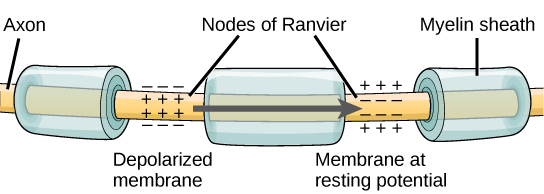
For an action potential to communicate information to another neuron, it must travel along the axon and reach the axon terminals where it can initiate neurotransmitter release. The speed of conduction of an action potential along an axon is influenced by both the diameter of the axon and the axon’s resistance to current leak. Myelin acts as an insulator that prevents current from leaving the axon; this increases the speed of action potential conduction. In demyelinating diseases like multiple sclerosis, action potential conduction slows because current leaks from previously insulated axon areas. The nodes of Ranvier, illustrated in [link] are gaps in the myelin sheath along the axon. These unmyelinated spaces are about one micrometer long and contain voltage gated Na + and K + channels. Flow of ions through these channels, particularly the Na + channels, regenerates the action potential over and over again along the axon. This ‘jumping’ of the action potential from one node to the next is called saltatory conduction    . If nodes of Ranvier were not present along an axon, the action potential would propagate very slowly since Na + and K + channels would have to continuously regenerate action potentials at every point along the axon instead of at specific points. Nodes of Ranvier also save energy for the neuron since the channels only need to be present at the nodes and not along the entire axon.

Illustration shows an axon covered in three bands of myelin sheath. Between the sheath coverings the axon is exposed. The uncovered parts of the axon are called nodes of Ranvier. In the illustration, the left node of Ranvier is depolarized such that the membrane potential is positive inside and negative outside. The right membrane of the right node is at the resting potential, negative inside and positive outside. An arrow indicates that the depolarization jumps from the left node to the right, so that the right node becomes depolarized.
Nodes of Ranvier are gaps in myelin coverage along axons. Nodes contain voltage-gated K + and Na + channels. Action potentials travel down the axon by jumping from one node to the next.

Synaptic transmission

The synapse or “gap” is the place where information is transmitted from one neuron to another. Synapses usually form between axon terminals and dendritic spines, but this is not universally true. There are also axon-to-axon, dendrite-to-dendrite, and axon-to-cell body synapses. The neuron transmitting the signal is called the presynaptic neuron, and the neuron receiving the signal is called the postsynaptic neuron. Note that these designations are relative to a particular synapse—most neurons are both presynaptic and postsynaptic. There are two types of synapses: chemical and electrical.

Chemical synapse

When an action potential reaches the axon terminal it depolarizes the membrane and opens voltage-gated Na + channels. Na + ions enter the cell, further depolarizing the presynaptic membrane. This depolarization causes voltage-gated Ca 2+ channels to open. Calcium ions entering the cell initiate a signaling cascade that causes small membrane-bound vesicles, called synaptic vesicles , containing neurotransmitter molecules to fuse with the presynaptic membrane. Synaptic vesicles are shown in [link] , which is an image from a scanning electron microscope.

The axon terminal is spherical. A section is sliced off, revealing small blue and orange vesicles just inside.
This pseudocolored image taken with a scanning electron microscope shows an axon terminal that was broken open to reveal synaptic vesicles (blue and orange) inside the neuron. (credit: modification of work by Tina Carvalho, NIH-NIGMS; scale-bar data from Matt Russell)

Questions & Answers

calculate molarity of NaOH solution when 25.0ml of NaOH titrated with 27.2ml of 0.2m H2SO4
Gasin Reply
what's Thermochemistry
rhoda Reply
the study of the heat energy which is associated with chemical reactions
Kaddija
How was CH4 and o2 was able to produce (Co2)and (H2o
Edafe Reply
explain please
Victory
First twenty elements with their valences
Martine Reply
what is chemistry
asue Reply
what is atom
asue
what is the best way to define periodic table for jamb
Damilola Reply
what is the change of matter from one state to another
Elijah Reply
what is isolation of organic compounds
IKyernum Reply
what is atomic radius
ThankGod Reply
Read Chapter 6, section 5
Dr
Read Chapter 6, section 5
Kareem
Atomic radius is the radius of the atom and is also called the orbital radius
Kareem
atomic radius is the distance between the nucleus of an atom and its valence shell
Amos
Read Chapter 6, section 5
paulino
Bohr's model of the theory atom
Ayom Reply
is there a question?
Dr
when a gas is compressed why it becomes hot?
ATOMIC
It has no oxygen then
Goldyei
read the chapter on thermochemistry...the sections on "PV" work and the First Law of Thermodynamics should help..
Dr
Which element react with water
Mukthar Reply
Mgo
Ibeh
an increase in the pressure of a gas results in the decrease of its
Valentina Reply
definition of the periodic table
Cosmos Reply
What is the lkenes
Da Reply
what were atoms composed of?
Moses Reply
Got questions? Join the online conversation and get instant answers!
Jobilize.com Reply

Get Jobilize Job Search Mobile App in your pocket Now!

Get it on Google Play Download on the App Store Now


Source:  OpenStax, Biology. OpenStax CNX. Feb 29, 2016 Download for free at http://cnx.org/content/col11448/1.10
Google Play and the Google Play logo are trademarks of Google Inc.

Notification Switch

Would you like to follow the 'Biology' conversation and receive update notifications?

Ask