<< Chapter < Page Chapter >> Page >
Photo (a) shows a plant with many fuzzy white hairs growing from its surface. Scanning electron micrograph (b) shows branched tree-like hairs emerging from the surface of a leaf. The trunk of each hair is about 250 microns tall. Branches are somewhat shorter. Scanning electron micrograph (c) shows many multi-pronged hairs about 100 microns long that look like sea anemones scattered across a leaf surface.
Trichomes give leaves a fuzzy appearance as in this (a) sundew ( Drosera sp.). Leaf trichomes include (b) branched trichomes on the leaf of Arabidopsis lyrata and (c) multibranched trichomes on a mature Quercus marilandica leaf. (credit a: John Freeland; credit b, c: modification of work by Robert R. Wise; scale-bar data from Matt Russell)

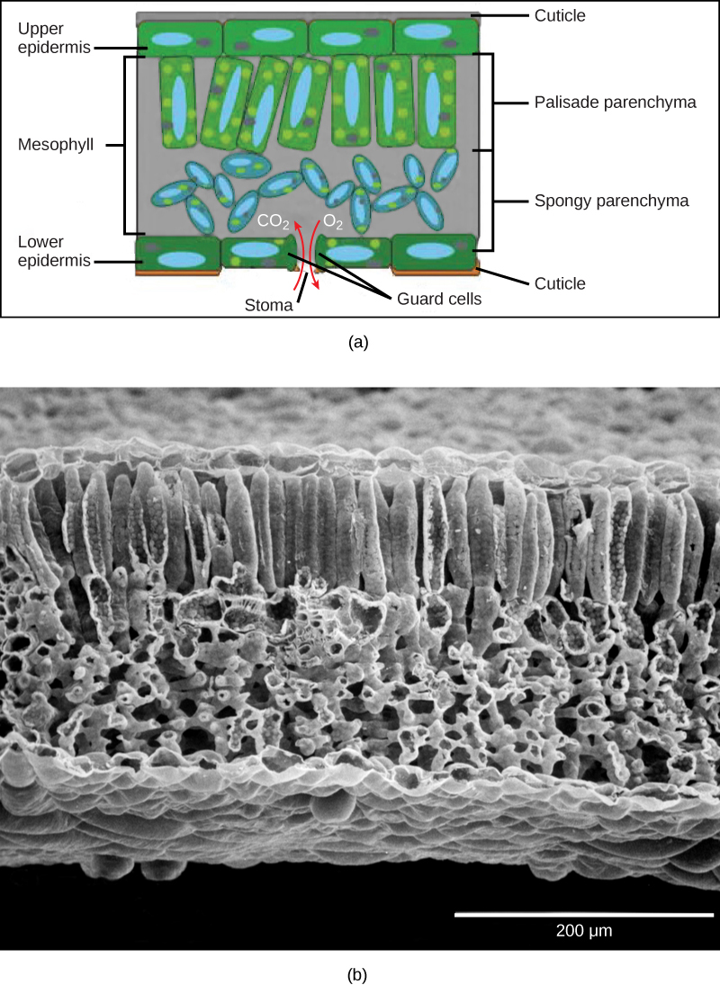
Below the epidermis of dicot leaves are layers of cells known as the mesophyll, or “middle leaf.” The mesophyll of most leaves typically contains two arrangements of parenchyma cells: the palisade parenchyma and spongy parenchyma ( [link] ). The palisade parenchyma (also called the palisade mesophyll) has column-shaped, tightly packed cells, and may be present in one, two, or three layers. Below the palisade parenchyma are loosely arranged cells of an irregular shape. These are the cells of the spongy parenchyma (or spongy mesophyll). The air space found between the spongy parenchyma cells allows gaseous exchange between the leaf and the outside atmosphere through the stomata. In aquatic plants, the intercellular spaces in the spongy parenchyma help the leaf float. Both layers of the mesophyll contain many chloroplasts. Guard cells are the only epidermal cells to contain chloroplasts.

 Part A is a leaf cross section illustration. A flat layer of rectangular cells make up the upper and lower epidermis. A cuticle layer protects the outside of both epidermal layers. A stomatal pore in the lower epidermis allows carbon dioxide to enter and oxygen to leave. Oval guard cells surround the pore. Sandwiched between the upper and lower epidermis is the mesophyll. The upper part of the mesophyll is comprised of columnar cells called palisade parenchyma. The lower part of the mesophyll is made up of loosely packed spongy parenchyma. Part B is a scanning electron micrograph of a leaf in which all the layers described above are visible. Palisade cells are about 50 microns tall and 10 microns wide and are covered with tiny bumps, which are the chloroplasts. Spongy cells smaller and irregularly shaped. Several large bumps about 20 microns across project from the lower surface of the leaf.
In the (a) leaf drawing, the central mesophyll is sandwiched between an upper and lower epidermis. The mesophyll has two layers: an upper palisade layer comprised of tightly packed, columnar cells, and a lower spongy layer, comprised of loosely packed, irregularly shaped cells. Stomata on the leaf underside allow gas exchange. A waxy cuticle covers all aerial surfaces of land plants to minimize water loss. These leaf layers are clearly visible in the (b) scanning electron micrograph. The numerous small bumps in the palisade parenchyma cells are chloroplasts. Chloroplasts are also present in the spongy parenchyma, but are not as obvious. The bumps protruding from the lower surface of the leave are glandular trichomes, which differ in structure from the stalked trichomes in [link] . (credit b: modification of work by Robert R. Wise)

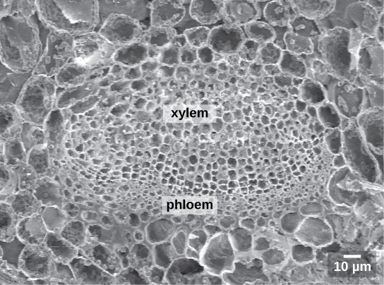
Like the stem, the leaf contains vascular bundles composed of xylem and phloem ( [link] ). The xylem consists of tracheids and vessels, which transport water and minerals to the leaves. The phloem transports the photosynthetic products from the leaf to the other parts of the plant. A single vascular bundle, no matter how large or small, always contains both xylem and phloem tissues.

 The scanning electron micrograph shows an oval vascular bundle. Small phloem cells make up the bottom of the bundle, and larger xylem cells make up the top. The bundle is surrounded by a ring of larger cells.
This scanning electron micrograph shows xylem and phloem in the leaf vascular bundle from the lyre-leaved sand cress ( Arabidopsis lyrata) . (credit: modification of work by Robert R. Wise; scale-bar data from Matt Russell)

Leaf adaptations

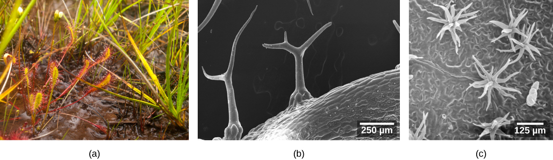
Coniferous plant species that thrive in cold environments, like spruce, fir, and pine, have leaves that are reduced in size and needle-like in appearance. These needle-like leaves have sunken stomata and a smaller surface area: two attributes that aid in reducing water loss. In hot climates, plants such as cacti have leaves that are reduced to spines, which in combination with their succulent stems, help to conserve water. Many aquatic plants have leaves with wide lamina that can float on the surface of the water, and a thick waxy cuticle on the leaf surface that repels water.

Questions & Answers

Discuss the differences between taste and flavor, including how other sensory inputs contribute to our  perception of flavor.
John Reply
taste refers to your understanding of the flavor . while flavor one The other hand is refers to sort of just a blend things.
Faith
While taste primarily relies on our taste buds, flavor involves a complex interplay between taste and aroma
Kamara
which drugs can we use for ulcers
Ummi Reply
omeprazole
Kamara
what
Renee
what is this
Renee
is a drug
Kamara
of anti-ulcer
Kamara
Omeprazole Cimetidine / Tagament For the complicated once ulcer - kit
Patrick
what is the function of lymphatic system
Nency Reply
Not really sure
Eli
to drain extracellular fluid all over the body.
asegid
The lymphatic system plays several crucial roles in the human body, functioning as a key component of the immune system and contributing to the maintenance of fluid balance. Its main functions include: 1. Immune Response: The lymphatic system produces and transports lymphocytes, which are a type of
asegid
to transport fluids fats proteins and lymphocytes to the blood stream as lymph
Adama
what is anatomy
Oyindarmola Reply
Anatomy is the identification and description of the structures of living things
Kamara
what's the difference between anatomy and physiology
Oyerinde Reply
Anatomy is the study of the structure of the body, while physiology is the study of the function of the body. Anatomy looks at the body's organs and systems, while physiology looks at how those organs and systems work together to keep the body functioning.
AI-Robot
what is enzymes all about?
Mohammed Reply
Enzymes are proteins that help speed up chemical reactions in our bodies. Enzymes are essential for digestion, liver function and much more. Too much or too little of a certain enzyme can cause health problems
Kamara
yes
Prince
how does the stomach protect itself from the damaging effects of HCl
Wulku Reply
little girl okay how does the stomach protect itself from the damaging effect of HCL
Wulku
it is because of the enzyme that the stomach produce that help the stomach from the damaging effect of HCL
Kamara
function of digestive system
Ali Reply
function of digestive
Ali
the diagram of the lungs
Adaeze Reply
what is the normal body temperature
Diya Reply
37 degrees selcius
Xolo
37°c
Stephanie
please why 37 degree selcius normal temperature
Mark
36.5
Simon
37°c
Iyogho
the normal temperature is 37°c or 98.6 °Fahrenheit is important for maintaining the homeostasis in the body the body regular this temperature through the process called thermoregulation which involves brain skin muscle and other organ working together to maintain stable internal temperature
Stephanie
37A c
Wulku
what is anaemia
Diya Reply
anaemia is the decrease in RBC count hemoglobin count and PVC count
Eniola
what is the pH of the vagina
Diya Reply
how does Lysin attack pathogens
Diya
acid
Mary
I information on anatomy position and digestive system and there enzyme
Elisha Reply
anatomy of the female external genitalia
Muhammad Reply
Organ Systems Of The Human Body (Continued) Organ Systems Of The Human Body (Continued)
Theophilus Reply
what's lochia albra
Kizito
Got questions? Join the online conversation and get instant answers!
Jobilize.com Reply

Get Jobilize Job Search Mobile App in your pocket Now!

Get it on Google Play Download on the App Store Now


Source:  OpenStax, Biology. OpenStax CNX. Feb 29, 2016 Download for free at http://cnx.org/content/col11448/1.10
Google Play and the Google Play logo are trademarks of Google Inc.

Notification Switch

Would you like to follow the 'Biology' conversation and receive update notifications?

Ask