<< Chapter < Page Chapter >> Page >
 Micrograph shows collenchyma cells, which are irregularly shaped and 25 to 50 microns across. The collenchyma cells are adjacent to a layer of rectangular cells that form the epidermis.
Collenchyma cell walls are uneven in thickness, as seen in this light micrograph. They provide support to plant structures. (credit: modification of work by Carl Szczerski; scale-bar data from Matt Russell)

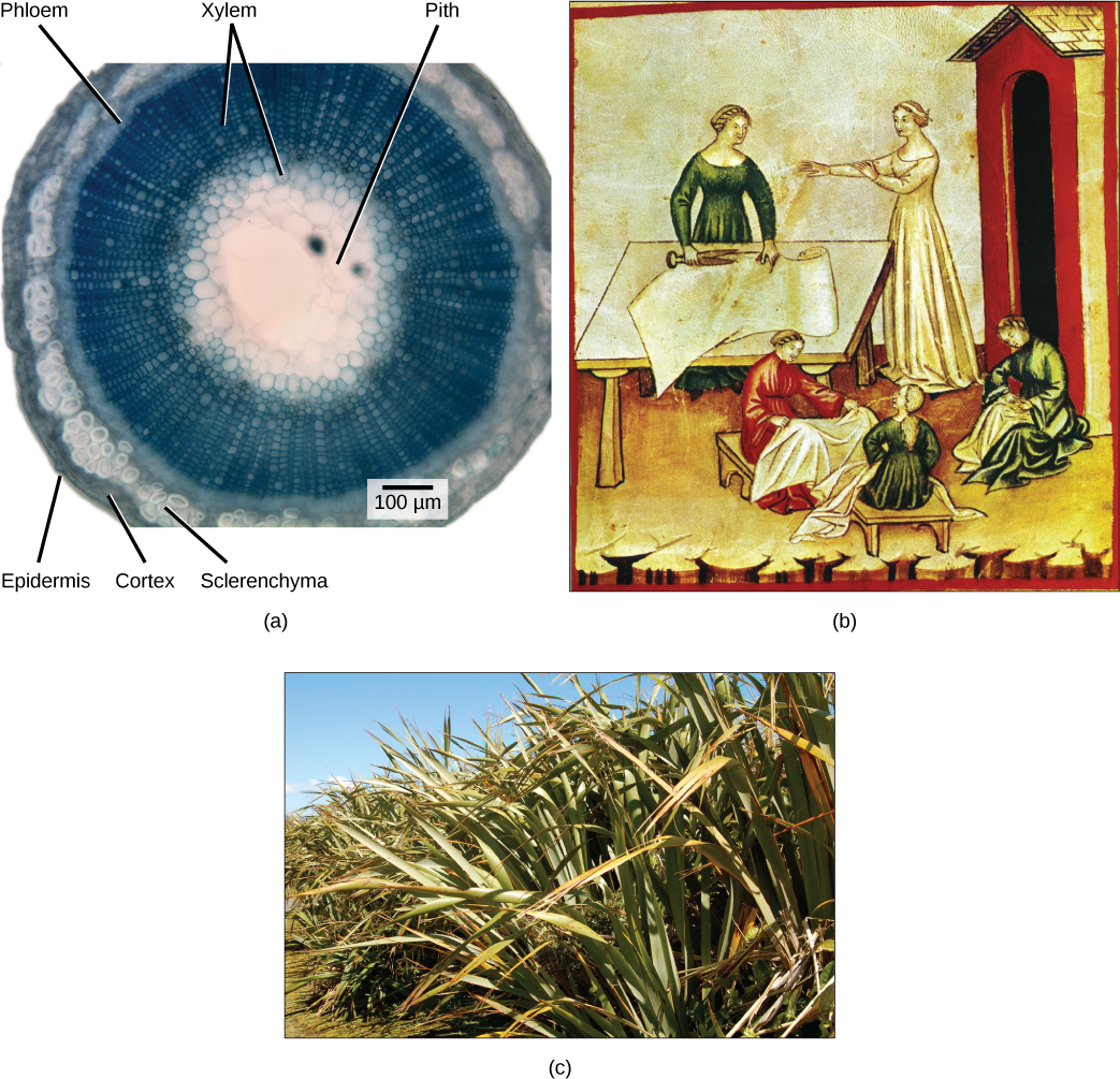
Sclerenchyma cells also provide support to the plant, but unlike collenchyma cells, many of them are dead at maturity. There are two types of sclerenchyma cells: fibers and sclereids. Both types have secondary cell walls that are thickened with deposits of lignin, an organic compound that is a key component of wood. Fibers are long, slender cells; sclereids are smaller-sized. Sclereids give pears their gritty texture. Humans use sclerenchyma fibers to make linen and rope ( [link] ).

Art connection

 Part A shows a cross section of a flax stem. The pith is white tissue in the center of the stem. Outside the pith is a layer of xylem. The inner xylem cells are large, while ones further out are smaller. The smaller xylem cells radiate out from the center, like spokes on a wheel. Outside the xylem is a ring of phloem cells. The phloem is surrounded by a layer of sclerenchyma cells, then a layer of cortex cells. Outside the cortex is the epidermis. Part B is a painting of women working with linen cloth. One is smoothing the cloth on a table, and the other women are sitting with linen on their laps. Part C is a photo of flax plants, which have long, wide leaves that taper toward narrow tips.
The central pith and outer cortex of the (a) flax stem are made up of parenchyma cells. Inside the cortex is a layer of sclerenchyma cells, which make up the fibers in flax rope and clothing. Humans have grown and harvested flax for thousands of years. In (b) this drawing, fourteenth-century women prepare linen. The (c) flax plant is grown and harvested for its fibers, which are used to weave linen, and for its seeds, which are the source of linseed oil. (credit a: modification of work by Emmanuel Boutet based on original work by Ryan R. MacKenzie; credit c: modification of work by Brian Dearth; scale-bar data from Matt Russell)

Which layers of the stem are made of parenchyma cells?

  1. cortex and pith
  2. phloem
  3. sclerenchyma
  4. xylem

Like the rest of the plant, the stem has three tissue systems: dermal, vascular, and ground tissue. Each is distinguished by characteristic cell types that perform specific tasks necessary for the plant’s growth and survival.

Dermal tissue

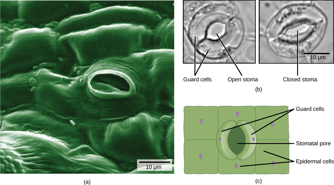
The dermal tissue of the stem consists primarily of epidermis    , a single layer of cells covering and protecting the underlying tissue. Woody plants have a tough, waterproof outer layer of cork cells commonly known as bark    , which further protects the plant from damage. Epidermal cells are the most numerous and least differentiated of the cells in the epidermis. The epidermis of a leaf also contains openings known as stomata, through which the exchange of gases takes place ( [link] ). Two cells, known as guard cells    , surround each leaf stoma, controlling its opening and closing and thus regulating the uptake of carbon dioxide and the release of oxygen and water vapor. Trichomes are hair-like structures on the epidermal surface. They help to reduce transpiration (the loss of water by aboveground plant parts), increase solar reflectance, and store compounds that defend the leaves against predation by herbivores.

 The electron micrograph in part A shows the lumpy, textured of a leaf epidermis. Individual cells look like pillows arranged side by side and fused together. In the center of the image is an oval pore about 10 microns across. Inside the pore, closed guard cells have the appearance of sealed lips. The two light micrographs in part B shows two kidney-shaped guard cells. In the left image, the stoma is open and round. In the right image, the stoma is closed and oval shaped. Part C is an illustration of the leaf epidermis with a oval stomatal pore in the center. Surrounding this pore are two kidney-shaped guard cells. Rectangular epidermal cells surround the guard cells.
Openings called stomata (singular: stoma) allow a plant to take up carbon dioxide and release oxygen and water vapor. The (a) colorized scanning-electron micrograph shows a closed stoma of a dicot. Each stoma is flanked by two guard cells that regulate its (b) opening and closing. The (c) guard cells sit within the layer of epidermal cells (credit a: modification of work by Louisa Howard, Rippel Electron Microscope Facility, Dartmouth College; credit b: modification of work by June Kwak, University of Maryland; scale-bar data from Matt Russell)

Questions & Answers

prostaglandin and fever
Maha Reply
Discuss the differences between taste and flavor, including how other sensory inputs contribute to our  perception of flavor.
John Reply
taste refers to your understanding of the flavor . while flavor one The other hand is refers to sort of just a blend things.
Faith
While taste primarily relies on our taste buds, flavor involves a complex interplay between taste and aroma
Kamara
which drugs can we use for ulcers
Ummi Reply
omeprazole
Kamara
what
Renee
what is this
Renee
is a drug
Kamara
of anti-ulcer
Kamara
Omeprazole Cimetidine / Tagament For the complicated once ulcer - kit
Patrick
what is the function of lymphatic system
Nency Reply
Not really sure
Eli
to drain extracellular fluid all over the body.
asegid
The lymphatic system plays several crucial roles in the human body, functioning as a key component of the immune system and contributing to the maintenance of fluid balance. Its main functions include: 1. Immune Response: The lymphatic system produces and transports lymphocytes, which are a type of
asegid
to transport fluids fats proteins and lymphocytes to the blood stream as lymph
Adama
what is anatomy
Oyindarmola Reply
Anatomy is the identification and description of the structures of living things
Kamara
what's the difference between anatomy and physiology
Oyerinde Reply
Anatomy is the study of the structure of the body, while physiology is the study of the function of the body. Anatomy looks at the body's organs and systems, while physiology looks at how those organs and systems work together to keep the body functioning.
AI-Robot
what is enzymes all about?
Mohammed Reply
Enzymes are proteins that help speed up chemical reactions in our bodies. Enzymes are essential for digestion, liver function and much more. Too much or too little of a certain enzyme can cause health problems
Kamara
yes
Prince
how does the stomach protect itself from the damaging effects of HCl
Wulku Reply
little girl okay how does the stomach protect itself from the damaging effect of HCL
Wulku
it is because of the enzyme that the stomach produce that help the stomach from the damaging effect of HCL
Kamara
function of digestive system
Ali Reply
function of digestive
Ali
the diagram of the lungs
Adaeze Reply
what is the normal body temperature
Diya Reply
37 degrees selcius
Xolo
37°c
Stephanie
please why 37 degree selcius normal temperature
Mark
36.5
Simon
37°c
Iyogho
the normal temperature is 37°c or 98.6 °Fahrenheit is important for maintaining the homeostasis in the body the body regular this temperature through the process called thermoregulation which involves brain skin muscle and other organ working together to maintain stable internal temperature
Stephanie
37A c
Wulku
what is anaemia
Diya Reply
anaemia is the decrease in RBC count hemoglobin count and PVC count
Eniola
what is the pH of the vagina
Diya Reply
how does Lysin attack pathogens
Diya
acid
Mary
I information on anatomy position and digestive system and there enzyme
Elisha Reply
anatomy of the female external genitalia
Muhammad Reply
Got questions? Join the online conversation and get instant answers!
Jobilize.com Reply

Get Jobilize Job Search Mobile App in your pocket Now!

Get it on Google Play Download on the App Store Now


Source:  OpenStax, Biology. OpenStax CNX. Feb 29, 2016 Download for free at http://cnx.org/content/col11448/1.10
Google Play and the Google Play logo are trademarks of Google Inc.

Notification Switch

Would you like to follow the 'Biology' conversation and receive update notifications?

Ask