<< Chapter < Page Chapter >> Page >

The unique sequence for every protein is ultimately determined by the gene encoding the protein. A change in nucleotide sequence of the gene’s coding region may lead to a different amino acid being added to the growing polypeptide chain, causing a change in protein structure and function. In sickle cell anemia, the hemoglobin β chain (a small portion of which is shown in [link] ) has a single amino acid change, out of 600 total amino acids, causing a change in protein structure and function.

A portion of the hemoglobin amino acid sequence is shown. The normal hemoglobin beta chain has a glutamate at position six. The sickle cell beta chain has a valine at this position.
The beta chain of hemoglobin is 147 amino acids in length, yet a single amino acid substitution leads to sickle cell anemia. In normal hemoglobin, the amino acid at position seven is glutamate. In sickle cell hemoglobin, this glutamate is replaced by a valine.

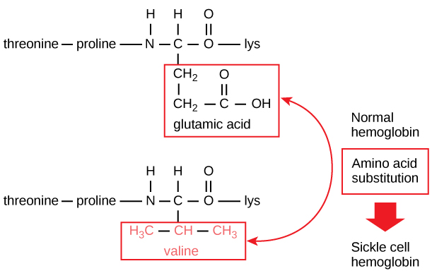
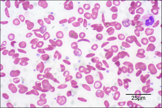
Because of this change of one amino acid in the chain, hemoglobin molecules form long fibers that distort the biconcave, or disc-shaped, red blood cells and assume a crescent or “sickle” shape, which clogs arteries ( [link] ). This can lead to myriad serious health problems such as breathlessness, dizziness, headaches, and abdominal pain for those affected by this disease.

This electron micrograph shows red blood cells from a patient with sickle cell anemia. Most of the cells have a normal, disk shape, but about one in five has a sickle shape. A normal blood cell is eight microns across.
In this blood smear, visualized at 535x magnification using bright field microscopy, sickle cells are crescent shaped, while normal cells are disc-shaped. (credit: modification of work by Ed Uthman; scale-bar data from Matt Russell)

Secondary structure

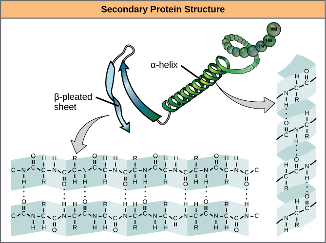
The local folding of the polypeptide in some regions gives rise to the secondary structure    of the protein. The most common are the α -helix and β -pleated sheet structures ( [link] ).

The illustration shows an alpha helix protein structure, which coils like a spring, and a beta-pleated sheet structure, which forms flat sheets stacked together. In an alpha-helix, hydrogen bonding occurs between the carbonyl group of one amino acid and the amino group of the amino acid that occurs four residues later. In a beta-pleated sheet, hydrogen bonding occurs between two different lengths of peptide that are antiparallel to one another.
The α -helix and β -pleated sheet are secondary structures of proteins that form because of hydrogen bonding between carbonyl and amino groups in the peptide backbone. Certain amino acids have a propensity to form an α -helix, while others have a propensity to form a β -pleated sheet.

Tertiary structure

The unique three-dimensional structure of a polypeptide is its tertiary structure    ( [link] ). This structure is in part due to chemical interactions at work on the polypeptide chain. Primarily, the interactions among R groups creates the complex three-dimensional tertiary structure of a protein. The nature of the R groups found in the amino acids involved can counteract the formation of the hydrogen bonds described for standard secondary structures. For example, R groups with like charges are repelled by each other and those with unlike charges are attracted to each other. When protein folding takes place, the hydrophobic R groups of nonpolar amino acids lay in the interior of the protein, whereas the hydrophilic R groups lay on the outside. The former types of interactions are also known as hydrophobic interactions. Interaction between cysteine side chains forms disulfide linkages in the presence of oxygen, the only covalent bond forming during protein folding.

This illustration shows a polypeptide backbone folded into a three-dimensional structure. Chemical interactions between amino acid side chains maintain its shape. These include an ionic bond between an amino group and a carboxyl group, hydrophobic interactions between two hydrophobic side chains, a hydrogen bond between a hydroxyl group and a carbonyl group, and a disulfide linkage.
The tertiary structure of proteins is determined by a variety of chemical interactions. These include hydrophobic interactions, ionic bonding, hydrogen bonding and disulfide linkages.

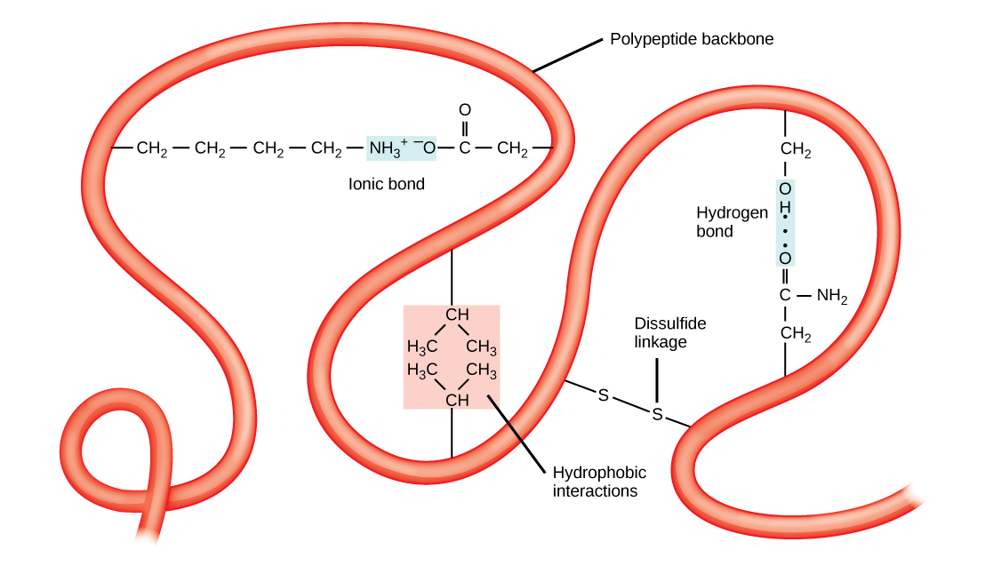
All of these interactions, weak and strong, determine the final three-dimensional shape of the protein. When a protein loses its three-dimensional shape, it may no longer be functional.

Get Jobilize Job Search Mobile App in your pocket Now!

Get it on Google Play Download on the App Store Now


Source:  OpenStax, General biology part i - mixed majors. OpenStax CNX. May 16, 2016 Download for free at http://legacy.cnx.org/content/col11749/1.5
Google Play and the Google Play logo are trademarks of Google Inc.

Notification Switch

Would you like to follow the 'General biology part i - mixed majors' conversation and receive update notifications?

Ask