<< Chapter < Page Chapter >> Page >

Art connection

Illustration shows the location of various mechanoreceptors in a cross section of the epidermis and dermis. A nerve runs along the middle of the dermis, and all the mechanoreceptors are connected to it. Ruffini endings, Merkel’s disks, and Meissner’s corpuscles are all located in the upper dermis above the nerve. Ruffini endings are bulbous, horizontal mechanoreceptors located in the middle of the upper dermis. Meissner’s corpuscles are bulbous, vertical mechanoreceptors that touch the bottom of the epidermis. Merkel’s disks have finger-like projections that also touch the bottom of the epidermis. The last type of mechanoreceptor, Pacini corpuscles, are oval mechanoreceptors located in the lower dermis.
Four of the primary mechanoreceptors in human skin are shown. Merkel’s disks, which are unencapsulated, respond to light touch. Meissner’s corpuscles, Ruffini endings, Pacinian corpuscles, and Krause end bulbs are all encapsulated. Meissner’s corpuscles respond to touch and low-frequency vibration. Ruffini endings detect stretch, deformation within joints, and warmth. Pacinian corpuscles detect transient pressure and high-frequency vibration. Krause end bulbs detect cold.

Which of the following statements about mechanoreceptors is false?

  1. Pacini corpuscles are found in both glabrous and hairy skin.
  2. Merkel’s disks are abundant on the fingertips and lips.
  3. Ruffini endings are encapsulated mechanoreceptors.
  4. Meissner’s corpuscles extend into the lower dermis.

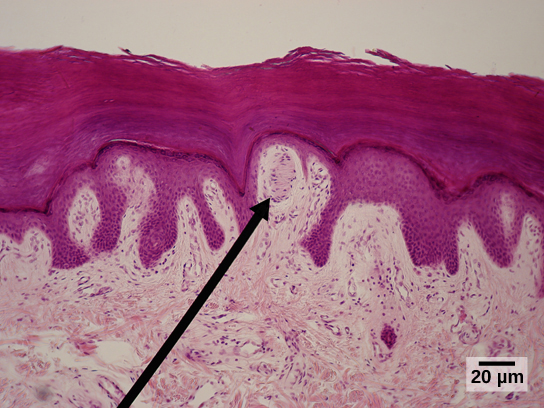
Meissner’s corpuscles , (shown in [link] ) also known as tactile corpuscles, are found in the upper dermis, but they project into the epidermis. They, too, are found primarily in the glabrous skin on the fingertips and eyelids. They respond to fine touch and pressure, but they also respond to low-frequency vibration or flutter. They are rapidly adapting, fluid-filled, encapsulated neurons with small, well-defined borders and are responsive to fine details. Like Merkel’s disks, Meissner’s corpuscles are not as plentiful in the palms as they are in the fingertips.

Micrograph shows the epidermis, which stains dark pink and the dermis, which stains light pink. Finger-like projections of epidermis extend into the dermis. Between two of these fingers is an oval Meissner corpuscle about ten microns across and 20 microns long.
Meissner corpuscles in the fingertips, such as the one viewed here using bright field light microscopy, allow for touch discrimination of fine detail. (credit: modification of work by "Wbensmith"/Wikimedia Commons; scale-bar data from Matt Russell)

Deeper in the epidermis, near the base, are Ruffini endings , which are also known as bulbous corpuscles. They are found in both glabrous and hairy skin. These are slow-adapting, encapsulated mechanoreceptors that detect skin stretch and deformations within joints, so they provide valuable feedback for gripping objects and controlling finger position and movement. Thus, they also contribute to proprioception and kinesthesia. Ruffini endings also detect warmth. Note that these warmth detectors are situated deeper in the skin than are the cold detectors. It is not surprising, then, that humans detect cold stimuli before they detect warm stimuli.

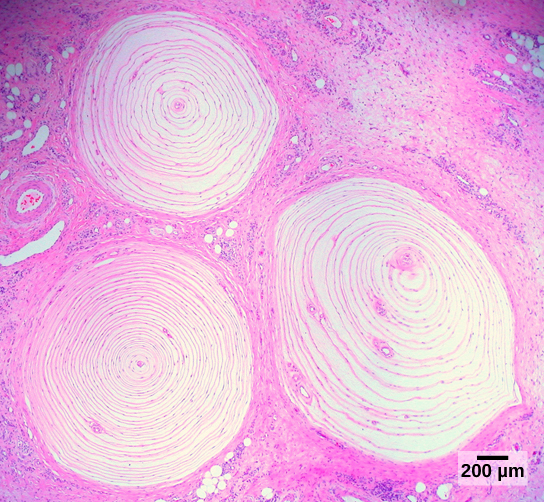
Pacinian corpuscles (seen in [link] ) are located deep in the dermis of both glabrous and hairy skin and are structurally similar to Meissner’s corpuscles; they are found in the bone periosteum, joint capsules, pancreas and other viscera, breast, and genitals. They are rapidly adapting mechanoreceptors that sense deep transient (but not prolonged) pressure and high-frequency vibration. Pacinian receptors detect pressure and vibration by being compressed, stimulating their internal dendrites. There are fewer Pacinian corpuscles and Ruffini endings in skin than there are Merkel’s disks and Meissner’s corpuscles.

Micrograph shows three Pacinian corpuscles embedded in the dermis. The corpuscles are round and about 1.4 millimeters across and have rings, like a tree stump.
Pacinian corpuscles, such as these visualized using bright field light microscopy, detect pressure (touch) and high-frequency vibration. (credit: modification of work by Ed Uthman; scale-bar data from Matt Russell)

Questions & Answers

what is biology
Hajah Reply
the study of living organisms and their interactions with one another and their environments
AI-Robot
what is biology
Victoria Reply
HOW CAN MAN ORGAN FUNCTION
Alfred Reply
the diagram of the digestive system
Assiatu Reply
allimentary cannel
Ogenrwot
How does twins formed
William Reply
They formed in two ways first when one sperm and one egg are splited by mitosis or two sperm and two eggs join together
Oluwatobi
what is genetics
Josephine Reply
Genetics is the study of heredity
Misack
how does twins formed?
Misack
What is manual
Hassan Reply
discuss biological phenomenon and provide pieces of evidence to show that it was responsible for the formation of eukaryotic organelles
Joseph Reply
what is biology
Yousuf Reply
the study of living organisms and their interactions with one another and their environment.
Wine
discuss the biological phenomenon and provide pieces of evidence to show that it was responsible for the formation of eukaryotic organelles in an essay form
Joseph Reply
what is the blood cells
Shaker Reply
list any five characteristics of the blood cells
Shaker
lack electricity and its more savely than electronic microscope because its naturally by using of light
Abdullahi Reply
advantage of electronic microscope is easily and clearly while disadvantage is dangerous because its electronic. advantage of light microscope is savely and naturally by sun while disadvantage is not easily,means its not sharp and not clear
Abdullahi
cell theory state that every organisms composed of one or more cell,cell is the basic unit of life
Abdullahi
is like gone fail us
DENG
cells is the basic structure and functions of all living things
Ramadan
What is classification
ISCONT Reply
is organisms that are similar into groups called tara
Yamosa
in what situation (s) would be the use of a scanning electron microscope be ideal and why?
Kenna Reply
A scanning electron microscope (SEM) is ideal for situations requiring high-resolution imaging of surfaces. It is commonly used in materials science, biology, and geology to examine the topography and composition of samples at a nanoscale level. SEM is particularly useful for studying fine details,
Hilary
cell is the building block of life.
Condoleezza Reply

Get Jobilize Job Search Mobile App in your pocket Now!

Get it on Google Play Download on the App Store Now


Source:  OpenStax, Biology. OpenStax CNX. Feb 29, 2016 Download for free at http://cnx.org/content/col11448/1.10
Google Play and the Google Play logo are trademarks of Google Inc.

Notification Switch

Would you like to follow the 'Biology' conversation and receive update notifications?

Ask