<< Chapter < Page Chapter >> Page >

Art connection

 Ascomycetes have both sexual and asexual life cycles. In the asexual life cycle, the haploid (1n) mycelium branches into a chain of cells called the conidiophore. Spores bud from the end of the conidiophore and germinate to form more mycelia. In the sexual life cycle, a round structure called an antheridium buds from the male strain, and a similar structure called the ascogonium buds from the female strain. In a process called plasmogamy, the ascogonium and antheridium fuse to form a cell with multiple haploid nuclei. Mitosis and cell division result in the growth of many hyphae, which form a fruiting body called the ascocarp. The hyphae are dikaryotic, meaning they have two haploid nuclei. Asci form at the tips of these hyphae. In a process called karyogamy, the nuclei in the asci fuse to form a diploid (2n) zygote. The zygote undergoes meiosis without cell division, resulting in an ascus with four 1n nuclei arranged in a row. Each nucleus undergoes mitosis, resulting in eight ascospores, which are also arranged in a row at the tip of the hyphae. Dispersal and germination results in the growth of new mycelia.
The lifecycle of an ascomycete is characterized by the production of asci during the sexual phase. The haploid phase is the predominant phase of the life cycle.

Which of the following statements is true?

  1. A dikaryotic ascus that forms in the ascocarp undergoes karyogamy, meiosis, and mitosis to form eight ascospores.
  2. A diploid ascus that forms in the ascocarp undergoes karyogamy, meiosis, and mitosis to form eight ascospores.
  3. A haploid zygote that forms in the ascocarp undergoes karyogamy, meiosis, and mitosis to form eight ascospores.
  4. A dikaryotic ascus that forms in the ascocarp undergoes plasmogamy, meiosis, and mitosis to form eight ascospores.
Micrograph shows asci, which appear as multiple, sphere-like shapes fused together into a structure about 7 microns across, and ascospores, which are small, light blue ovals about two microns wide by three microns long released from the asci.
The bright field light micrograph shows ascospores being released from asci in the fungus Talaromyces flavus var. flavus . (credit: modification of work by Dr. Lucille Georg, CDC; scale-bar data from Matt Russell)

Basidiomycota: the club fungi

The fungi in the Phylum Basidiomycota    are easily recognizable under a light microscope by their club-shaped fruiting bodies called basidia (singular, basidium    ), which are the swollen terminal cell of a hypha. The basidia, which are the reproductive organs of these fungi, are often contained within the familiar mushroom, commonly seen in fields after rain, on the supermarket shelves, and growing on your lawn ( [link] ). These mushroom-producing basidiomyces are sometimes referred to as “gill fungi” because of the presence of gill-like structures on the underside of the cap. The “gills” are actually compacted hyphae on which the basidia are borne. This group also includes shelf fungus, which cling to the bark of trees like small shelves. In addition, the basidiomycota includes smuts and rusts, which are important plant pathogens; toadstools, and shelf fungi stacked on tree trunks. Most edible fungi belong to the Phylum Basidiomycota; however, some basidiomycetes produce deadly toxins. For example, Cryptococcus neoformans causes severe respiratory illness.

 Photo shows toadstools growing in a ring on a lawn.
The fruiting bodies of a basidiomycete form a ring in a meadow, commonly called “fairy ring.” The best-known fairy ring fungus has the scientific name Marasmius oreades . The body of this fungus, its mycelium, is underground and grows outward in a circle. As it grows, the mycelium depletes the soil of nitrogen, causing the mycelia to grow away from the center and leading to the “fairy ring” of fruiting bodies where there is adequate soil nitrogen. (Credit: "Cropcircles"/Wikipedia Commons)]

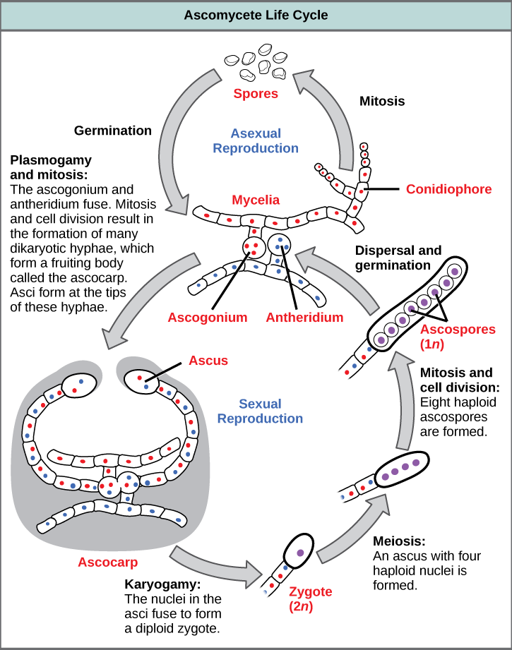
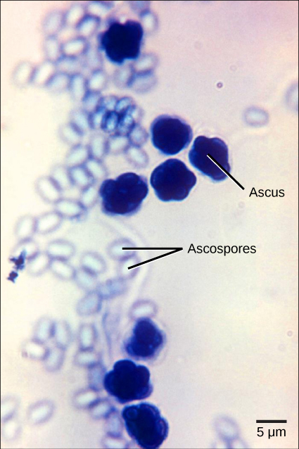
The lifecycle of basidiomycetes includes alternation of generations ( [link] ). Spores are generally produced through sexual reproduction, rather than asexual reproduction. The club-shaped basidium carries spores called basidiospores. In the basidium, nuclei of two different mating strains fuse (karyogamy), giving rise to a diploid zygote that then undergoes meiosis. The haploid nuclei migrate into basidiospores, which germinate and generate monokaryotic hyphae. The mycelium that results is called a primary mycelium. Mycelia of different mating strains can combine and produce a secondary mycelium that contains haploid nuclei of two different mating strains. This is the dikaryotic stage of the basidiomyces lifecyle and and it is the dominant stage. Eventually, the secondary mycelium generates a basidiocarp    , which is a fruiting body that protrudes from the ground—this is what we think of as a mushroom. The basidiocarp bears the developing basidia on the gills under its cap.

Questions & Answers

what is biology
Hajah Reply
the study of living organisms and their interactions with one another and their environments
AI-Robot
what is biology
Victoria Reply
HOW CAN MAN ORGAN FUNCTION
Alfred Reply
the diagram of the digestive system
Assiatu Reply
allimentary cannel
Ogenrwot
How does twins formed
William Reply
They formed in two ways first when one sperm and one egg are splited by mitosis or two sperm and two eggs join together
Oluwatobi
what is genetics
Josephine Reply
Genetics is the study of heredity
Misack
how does twins formed?
Misack
What is manual
Hassan Reply
discuss biological phenomenon and provide pieces of evidence to show that it was responsible for the formation of eukaryotic organelles
Joseph Reply
what is biology
Yousuf Reply
the study of living organisms and their interactions with one another and their environment.
Wine
discuss the biological phenomenon and provide pieces of evidence to show that it was responsible for the formation of eukaryotic organelles in an essay form
Joseph Reply
what is the blood cells
Shaker Reply
list any five characteristics of the blood cells
Shaker
lack electricity and its more savely than electronic microscope because its naturally by using of light
Abdullahi Reply
advantage of electronic microscope is easily and clearly while disadvantage is dangerous because its electronic. advantage of light microscope is savely and naturally by sun while disadvantage is not easily,means its not sharp and not clear
Abdullahi
cell theory state that every organisms composed of one or more cell,cell is the basic unit of life
Abdullahi
is like gone fail us
DENG
cells is the basic structure and functions of all living things
Ramadan
What is classification
ISCONT Reply
is organisms that are similar into groups called tara
Yamosa
in what situation (s) would be the use of a scanning electron microscope be ideal and why?
Kenna Reply
A scanning electron microscope (SEM) is ideal for situations requiring high-resolution imaging of surfaces. It is commonly used in materials science, biology, and geology to examine the topography and composition of samples at a nanoscale level. SEM is particularly useful for studying fine details,
Hilary
cell is the building block of life.
Condoleezza Reply

Get Jobilize Job Search Mobile App in your pocket Now!

Get it on Google Play Download on the App Store Now


Source:  OpenStax, Biology. OpenStax CNX. Feb 29, 2016 Download for free at http://cnx.org/content/col11448/1.10
Google Play and the Google Play logo are trademarks of Google Inc.

Notification Switch

Would you like to follow the 'Biology' conversation and receive update notifications?

Ask