<< Chapter < Page Chapter >> Page >

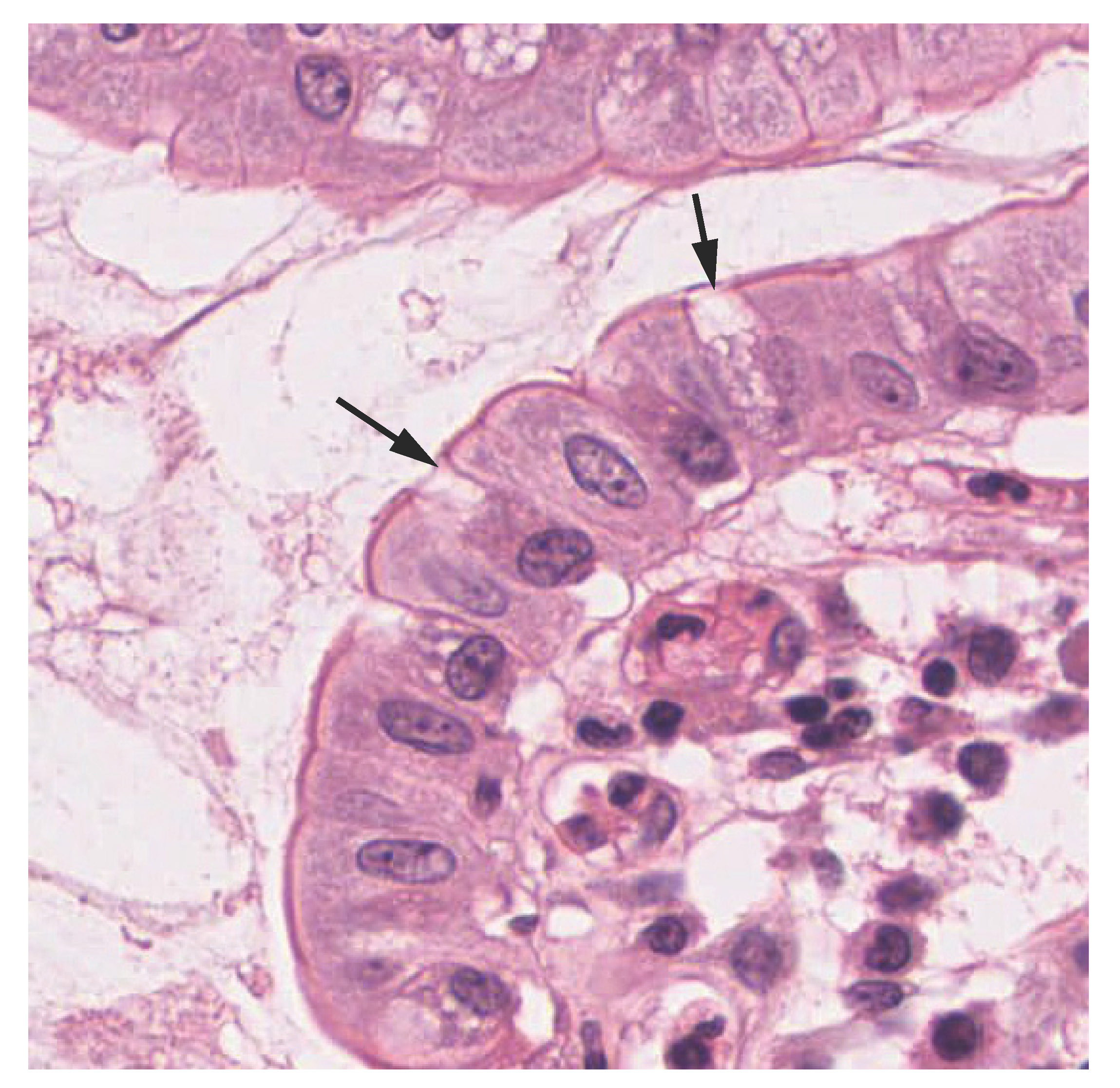
Pseudostratified columnar epithelium is a type of epithelium that appears to be stratified but instead consists of a single layer of irregularly shaped and differently sized columnar cells. In pseudostratified epithelium, nuclei of neighboring cells appear at different levels rather than clustered in the basal end. The arrangement gives the appearance of stratification; but in fact all the cells are in contact with the basal lamina, although some do not reach the apical surface. Pseudostratified columnar epithelium is found in the respiratory tract, where some of these cells have cilia.

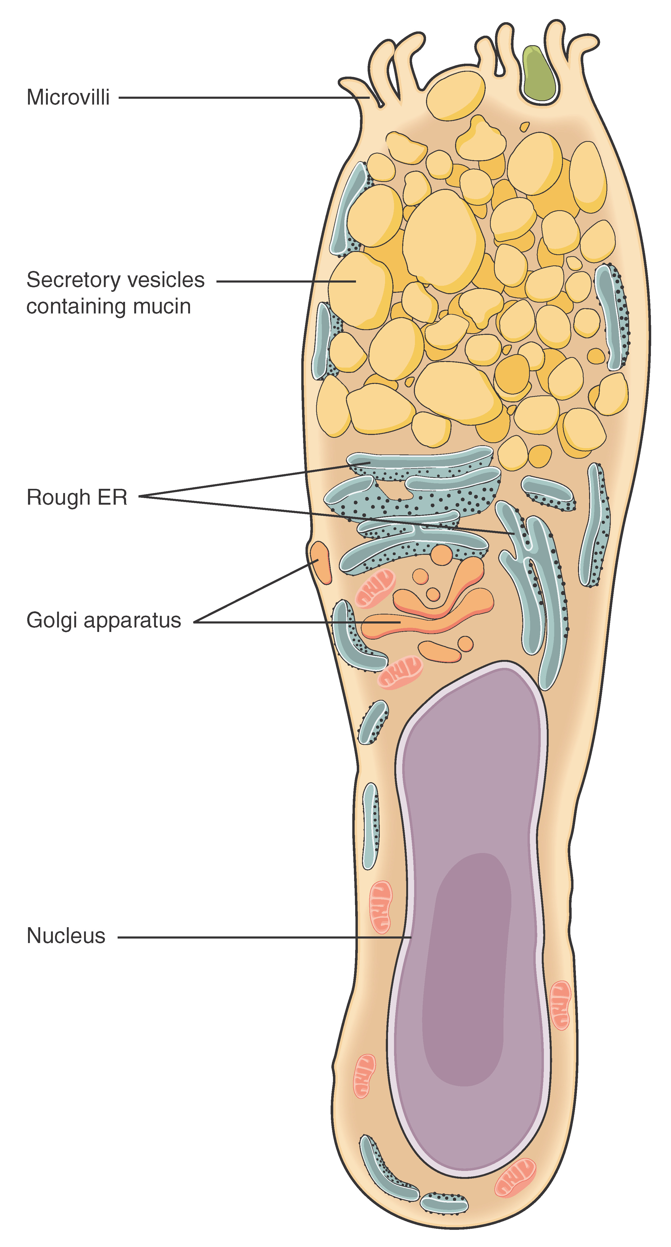
Both simple and pseudostratified columnar epithelia are heterogeneous epithelia because they include additional types of cells interspersed among the epithelial cells. For example, a goblet cell    is a mucous-secreting unicellular “gland” interspersed between the columnar epithelial cells of mucous membranes ( [link] ).

Goblet cell

This illustration shows a diagram of a goblet cell. The goblet cell is shaped roughly like an upside down vase. The enlarged end at the top contains six finger like projections labeled microvilli. Between the microvilli, secretary vesicles containing mucin are moving from the upper half of the cell toward the microvilli. Below the secretory vesicles are several rough endoplasmic reticula and an irregularly shaped Golgi apparatus with secretory vesicles budding off of it. The narrow, lower half of the cell contains the oval-shaped nucleus as well as a few mitochondria and segments of the endoplasmic reticulum. The second image is a micrograph of the innermost lining of the small intestine. This innermost lining is a simple columnar epithelium, with a single layer of rectangular cells oriented in a line. Occasionally, the line of epithelial cells is interrupted by a goblet cell. Goblet cells are thinner than the epithelial cells and appear roughly pill shaped. In this micrograph, the cells did not stain as darkly as the epithelial cells.
(a) In the lining of the small intestine, columnar epithelium cells are interspersed with goblet cells. (b) The arrows in this micrograph point to the mucous-secreting goblet cells. LM × 1600. (Micrograph provided by the Regents of University of Michigan Medical School © 2012)

View the University of Michigan WebScope at (External Link) to explore the tissue sample in greater detail.

Stratified epithelium

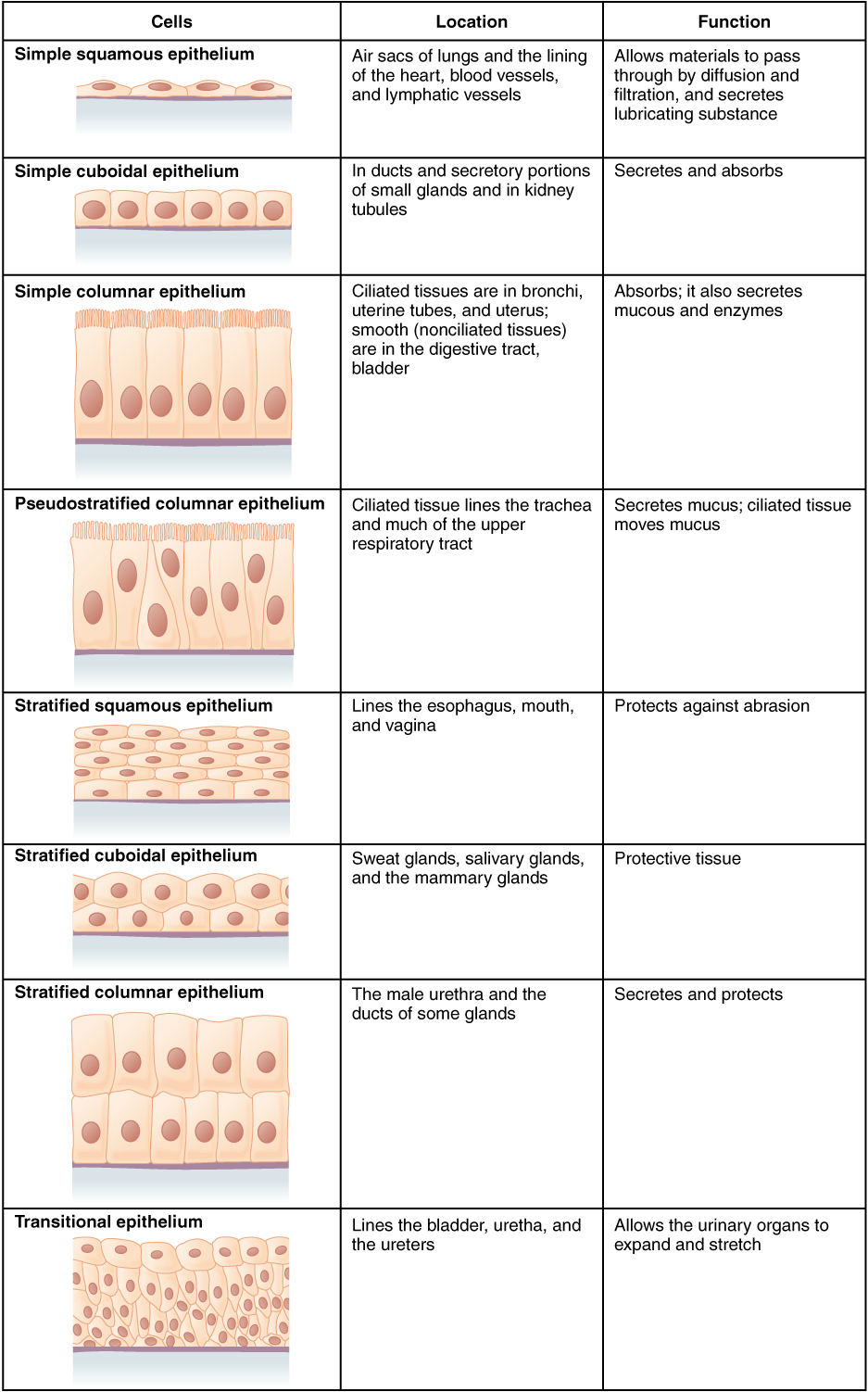
A stratified epithelium consists of several stacked layers of cells. This epithelium protects against physical and chemical wear and tear. The stratified epithelium is named by the shape of the most apical layer of cells, closest to the free space. Stratified squamous epithelium is the most common type of stratified epithelium in the human body. The apical cells are squamous, whereas the basal layer contains either columnar or cuboidal cells. The top layer may be covered with dead cells filled with keratin. Mammalian skin is an example of this dry, keratinized, stratified squamous epithelium. The lining of the mouth cavity is an example of an unkeratinized, stratified squamous epithelium. Stratified cuboidal epithelium and stratified columnar epithelium    can also be found in certain glands and ducts, but are uncommon in the human body.

Another kind of stratified epithelium is transitional epithelium    , so-called because of the gradual changes in the shapes of the apical cells as the bladder fills with urine. It is found only in the urinary system, specifically the ureters and urinary bladder. When the bladder is empty, this epithelium is convoluted and has cuboidal apical cells with convex, umbrella shaped, apical surfaces. As the bladder fills with urine, this epithelium loses its convolutions and the apical cells transition from cuboidal to squamous. It appears thicker and more multi-layered when the bladder is empty, and more stretched out and less stratified when the bladder is full and distended. [link] summarizes the different categories of epithelial cell tissue cells.

Summary of epithelial tissue cells

This figure is a table with three columns and eight rows. The leftmost column is titled cells, and contains a drawing in each row showing how epithelial cells are arranged above a basement membrane. The middle column is titled location, while the rightmost column is titled function. In a simple squamous epithelium, the cells are flattened and single-layered. Simple squamous cells are found in the air sacs of the lungs, in the lining of the heart, blood vessels and lymphatic vessels. Their function is to allow materials to pass through by diffusion and filtration, as well as to secrete lubricating substances. In a simple cuboidal epithelium, the cells are cube shaped and single layered and located in ducts and secretory portions of small glands as well as in the kidney tubules. The function of simple cuboidal epithelium is to secrete and absorb. In a simple columnar epithelium, the cells are rectangular and are attached to the basement membrane on one of their narrow sides, so that each cell is standing up like a column. There is only one layer of cells. Simple columnar epithelium is found in ciliated tissues including the bronchi, uterine tubes, and uterus, as well as in smooth, nonciliated tissues such as the digestive tract bladder. The function of simple columnar epithelium is to absorb substances but also to secrete mucous and enzymes. In a pseudostratified columnar epithelium, the cells are column-like in appearance, but they vary in height. The taller cells bend over the tops of the shorter cells so that the top of the epithelial tissue is continuous. There is only one layer of cells. Pseudostratified columnar epithelium lines the trachea and much of the upper respiratory tract. The function of pseudostratified columnar epithelium is to secrete mucous and also move that mucus using the hair like cilia projecting from the top of each cell. A stratified squamous epithelium contains many layers of flattened cells. Stratified squamous epithelium lines the esophagus, mouth, and vagina. The function of stratified squamous epithelium is to protect against abrasion. Stratified cuboidal epithelium contains many layers of cube-shaped cells. Stratified cuboidal epithelium is found in the sweat glands, salivary glands, and mammary glands. The function of stratified cuboidal epithelium is to protect other tissues of the body. Stratified columnar epithelium contains many layers of rectangular, column-shaped cells. Stratified columnar epithelium is located in the male urethra and the ducts of some glands. The function of stratified columnar epithelium is to secrete and protect. Transitional epithelium consists of many layers of irregularly shaped cells with diverse sizes. Transitional epithelium is found lining the bladder, urethra and ureters. The function of transitional epithelium is to allow the urinary organs to expand and stretch.

Watch this video to find out more about the anatomy of epithelial tissues. Where in the body would one find non-keratinizing stratified squamous epithelium?

Questions & Answers

how does Neisseria cause meningitis
Nyibol Reply
what is microbiologist
Muhammad Reply
what is errata
Muhammad
is the branch of biology that deals with the study of microorganisms.
Ntefuni Reply
What is microbiology
Mercy Reply
studies of microbes
Louisiaste
when we takee the specimen which lumbar,spin,
Ziyad Reply
How bacteria create energy to survive?
Muhamad Reply
Bacteria doesn't produce energy they are dependent upon their substrate in case of lack of nutrients they are able to make spores which helps them to sustain in harsh environments
_Adnan
But not all bacteria make spores, l mean Eukaryotic cells have Mitochondria which acts as powerhouse for them, since bacteria don't have it, what is the substitution for it?
Muhamad
they make spores
Louisiaste
what is sporadic nd endemic, epidemic
Aminu Reply
the significance of food webs for disease transmission
Abreham
food webs brings about an infection as an individual depends on number of diseased foods or carriers dully.
Mark
explain assimilatory nitrate reduction
Esinniobiwa Reply
Assimilatory nitrate reduction is a process that occurs in some microorganisms, such as bacteria and archaea, in which nitrate (NO3-) is reduced to nitrite (NO2-), and then further reduced to ammonia (NH3).
Elkana
This process is called assimilatory nitrate reduction because the nitrogen that is produced is incorporated in the cells of microorganisms where it can be used in the synthesis of amino acids and other nitrogen products
Elkana
Examples of thermophilic organisms
Shu Reply
Give Examples of thermophilic organisms
Shu
advantages of normal Flora to the host
Micheal Reply
Prevent foreign microbes to the host
Abubakar
they provide healthier benefits to their hosts
ayesha
They are friends to host only when Host immune system is strong and become enemies when the host immune system is weakened . very bad relationship!
Mark
what is cell
faisal Reply
cell is the smallest unit of life
Fauziya
cell is the smallest unit of life
Akanni
ok
Innocent
cell is the structural and functional unit of life
Hasan
is the fundamental units of Life
Musa
what are emergency diseases
Micheal Reply
There are nothing like emergency disease but there are some common medical emergency which can occur simultaneously like Bleeding,heart attack,Breathing difficulties,severe pain heart stock.Hope you will get my point .Have a nice day ❣️
_Adnan
define infection ,prevention and control
Innocent
I think infection prevention and control is the avoidance of all things we do that gives out break of infections and promotion of health practices that promote life
Lubega
Heyy Lubega hussein where are u from?
_Adnan
en français
Adama
which site have a normal flora
ESTHER Reply
Many sites of the body have it Skin Nasal cavity Oral cavity Gastro intestinal tract
Safaa
skin
Asiina
skin,Oral,Nasal,GIt
Sadik
How can Commensal can Bacteria change into pathogen?
Sadik
How can Commensal Bacteria change into pathogen?
Sadik
all
Tesfaye
by fussion
Asiina
what are the advantages of normal Flora to the host
Micheal
what are the ways of control and prevention of nosocomial infection in the hospital
Micheal
what is inflammation
Shelly Reply
part of a tissue or an organ being wounded or bruised.
Wilfred
what term is used to name and classify microorganisms?
Micheal Reply
Binomial nomenclature
adeolu
Got questions? Join the online conversation and get instant answers!
Jobilize.com Reply

Get Jobilize Job Search Mobile App in your pocket Now!

Get it on Google Play Download on the App Store Now


Source:  OpenStax, Anatomy & Physiology. OpenStax CNX. Feb 04, 2016 Download for free at http://legacy.cnx.org/content/col11496/1.8
Google Play and the Google Play logo are trademarks of Google Inc.

Notification Switch

Would you like to follow the 'Anatomy & Physiology' conversation and receive update notifications?

Ask