The brain case consists of eight bones. These include the paired parietal and temporal bones, plus the unpaired frontal, occipital, sphenoid, and ethmoid bones.
Parietal bone
The
parietal bone forms most of the upper lateral side of the skull (see
[link] ). These are paired bones, with the right and left parietal bones joining together at the top of the skull. Each parietal bone is also bounded anteriorly by the frontal bone, inferiorly by the temporal bone, and posteriorly by the occipital bone.
Temporal bone
The
temporal bone forms the lower lateral side of the skull (see
[link] ). Common wisdom has it that the temporal bone (temporal = “time”) is so named because this area of the head (the temple) is where hair typically first turns gray, indicating the passage of time.
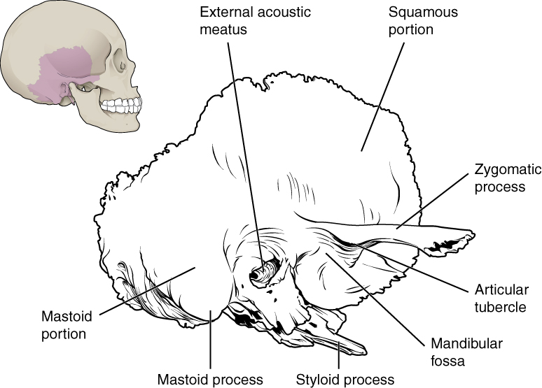
The temporal bone is subdivided into several regions (
[link] ). The flattened, upper portion is the squamous portion of the temporal bone. Below this area and projecting anteriorly is the zygomatic process of the temporal bone, which forms the posterior portion of the zygomatic arch. Posteriorly is the mastoid portion of the temporal bone. Projecting inferiorly from this region is a large prominence, the
mastoid process , which serves as a muscle attachment site. The mastoid process can easily be felt on the side of the head just behind your earlobe. On the interior of the skull, the petrous portion of each temporal bone forms the prominent, diagonally oriented
petrous ridge in the floor of the cranial cavity. Located inside each petrous ridge are small cavities that house the structures of the middle and inner ears.
Temporal bone
A lateral view of the isolated temporal bone shows the squamous, mastoid, and zygomatic portions of the temporal bone.
Important landmarks of the temporal bone, as shown in
[link] , include the following:
External acoustic meatus (ear canal)—This is the large opening on the lateral side of the skull that is associated with the ear.
Internal acoustic meatus —This opening is located inside the cranial cavity, on the medial side of the petrous ridge. It connects to the middle and inner ear cavities of the temporal bone.
Mandibular fossa —This is the deep, oval-shaped depression located on the external base of the skull, just in front of the external acoustic meatus. The mandible (lower jaw) joins with the skull at this site as part of the temporomandibular joint, which allows for movements of the mandible during opening and closing of the mouth.
Articular tubercle —The smooth ridge located immediately anterior to the mandibular fossa. Both the articular tubercle and mandibular fossa contribute to the temporomandibular joint, the joint that provides for movements between the temporal bone of the skull and the mandible.
Styloid process —Posterior to the mandibular fossa on the external base of the skull is an elongated, downward bony projection called the styloid process, so named because of its resemblance to a stylus (a pen or writing tool). This structure serves as an attachment site for several small muscles and for a ligament that supports the hyoid bone of the neck. (See also
[link] .)
Stylomastoid foramen —This small opening is located between the styloid process and mastoid process. This is the point of exit for the cranial nerve that supplies the facial muscles.
Carotid canal —The carotid canal is a zig-zag shaped tunnel that provides passage through the base of the skull for one of the major arteries that supplies the brain. Its entrance is located on the outside base of the skull, anteromedial to the styloid process. The canal then runs anteromedially within the bony base of the skull, and then turns upward to its exit in the floor of the middle cranial cavity, above the foramen lacerum.
the study of living organisms and their interactions with one another and their environment.
Wine
discuss the biological phenomenon and provide pieces of evidence to show that it was responsible for the formation of eukaryotic organelles in an essay form
advantage of electronic microscope is easily and clearly while disadvantage is dangerous because its electronic. advantage of light microscope is savely and naturally by sun while disadvantage is not easily,means its not sharp and not clear
Abdullahi
cell theory state that every organisms composed of one or more cell,cell is the basic unit of life
Abdullahi
is like gone fail us
DENG
cells is the basic structure and functions of all living things
A scanning electron microscope (SEM) is ideal for situations requiring high-resolution imaging of surfaces. It is commonly used in materials science, biology, and geology to examine the topography and composition of samples at a nanoscale level. SEM is particularly useful for studying fine details,