<< Chapter < Page Chapter >> Page >

Refinements and enhancements of X-ray techniques have continued throughout the twentieth and twenty-first centuries. Although often supplanted by more sophisticated imaging techniques, the X-ray remains a “workhorse” in medical imaging, especially for viewing fractures and for dentistry. The disadvantage of irradiation to the patient and the operator is now attenuated by proper shielding and by limiting exposure.

Modern medical imaging

X-rays can depict a two-dimensional image of a body region, and only from a single angle. In contrast, more recent medical imaging technologies produce data that is integrated and analyzed by computers to produce three-dimensional images or images that reveal aspects of body functioning.

Computed tomography

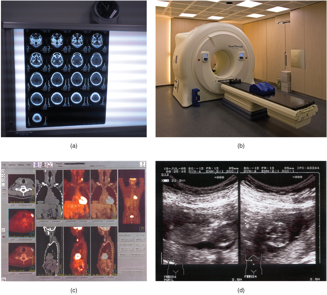
Tomography refers to imaging by sections. Computed tomography (CT) is a noninvasive imaging technique that uses computers to analyze several cross-sectional X-rays in order to reveal minute details about structures in the body ( [link] a ). The technique was invented in the 1970s and is based on the principle that, as X-rays pass through the body, they are absorbed or reflected at different levels. In the technique, a patient lies on a motorized platform while a computerized axial tomography (CAT) scanner rotates 360 degrees around the patient, taking X-ray images. A computer combines these images into a two-dimensional view of the scanned area, or “slice.”

Medical imaging techniques

These photos shows four types of imaging equipment. Photo A, the results of a CT scan, shows 17 different transverse views of the skull, each taken at a different depth along the superior-inferior axis. The images are translucent, similar to an X ray, and are viewed on a light board. Photo B shows an MRI machine, which is a large drum into which lying patients enter via a conveyor belt. Photo C shows computer images of the body taken with PET scans. This produces anterior, lateral, posterior, and transverse views of the body that reveal the structure of the internal organs. Photo D shows an ultrasound readout, which is black and white. The image depicts solid tissues as light areas and empty space as dark areas. Some of the features of a young fetus can be seen in the empty space at the center of the image. The space containing the fetus is surrounded by the solid tissue of the uterus.
(a) The results of a CT scan of the head are shown as successive transverse sections. (b) An MRI machine generates a magnetic field around a patient. (c) PET scans use radiopharmaceuticals to create images of active blood flow and physiologic activity of the organ or organs being targeted. (d) Ultrasound technology is used to monitor pregnancies because it is the least invasive of imaging techniques and uses no electromagnetic radiation. (credit a: Akira Ohgaki/flickr; credit b: “Digital Cate”/flickr; credit c: “Raziel”/Wikimedia Commons; credit d: “Isis”/Wikimedia Commons)

Since 1970, the development of more powerful computers and more sophisticated software has made CT scanning routine for many types of diagnostic evaluations. It is especially useful for soft tissue scanning, such as of the brain and the thoracic and abdominal viscera. Its level of detail is so precise that it can allow physicians to measure the size of a mass down to a millimeter. The main disadvantage of CT scanning is that it exposes patients to a dose of radiation many times higher than that of X-rays. In fact, children who undergo CT scans are at increased risk of developing cancer, as are adults who have multiple CT scans.

A CT or CAT scan relies on a circling scanner that revolves around the patient’s body. Watch this video to learn more about CT and CAT scans. What type of radiation does a CT scanner use?

Magnetic resonance imaging

Magnetic resonance imaging (MRI) is a noninvasive medical imaging technique based on a phenomenon of nuclear physics discovered in the 1930s, in which matter exposed to magnetic fields and radio waves was found to emit radio signals. In 1970, a physician and researcher named Raymond Damadian noticed that malignant (cancerous) tissue gave off different signals than normal body tissue. He applied for a patent for the first MRI scanning device, which was in use clinically by the early 1980s. The early MRI scanners were crude, but advances in digital computing and electronics led to their advancement over any other technique for precise imaging, especially to discover tumors. MRI also has the major advantage of not exposing patients to radiation.

Questions & Answers

what is phylogeny
Odigie Reply
evolutionary history and relationship of an organism or group of organisms
AI-Robot
ok
Deng
what is biology
Hajah Reply
the study of living organisms and their interactions with one another and their environments
AI-Robot
what is biology
Victoria Reply
HOW CAN MAN ORGAN FUNCTION
Alfred Reply
the diagram of the digestive system
Assiatu Reply
allimentary cannel
Ogenrwot
How does twins formed
William Reply
They formed in two ways first when one sperm and one egg are splited by mitosis or two sperm and two eggs join together
Oluwatobi
what is genetics
Josephine Reply
Genetics is the study of heredity
Misack
how does twins formed?
Misack
What is manual
Hassan Reply
discuss biological phenomenon and provide pieces of evidence to show that it was responsible for the formation of eukaryotic organelles
Joseph Reply
what is biology
Yousuf Reply
the study of living organisms and their interactions with one another and their environment.
Wine
discuss the biological phenomenon and provide pieces of evidence to show that it was responsible for the formation of eukaryotic organelles in an essay form
Joseph Reply
what is the blood cells
Shaker Reply
list any five characteristics of the blood cells
Shaker
lack electricity and its more savely than electronic microscope because its naturally by using of light
Abdullahi Reply
advantage of electronic microscope is easily and clearly while disadvantage is dangerous because its electronic. advantage of light microscope is savely and naturally by sun while disadvantage is not easily,means its not sharp and not clear
Abdullahi
cell theory state that every organisms composed of one or more cell,cell is the basic unit of life
Abdullahi
is like gone fail us
DENG
cells is the basic structure and functions of all living things
Ramadan
What is classification
ISCONT Reply
is organisms that are similar into groups called tara
Yamosa
in what situation (s) would be the use of a scanning electron microscope be ideal and why?
Kenna Reply
A scanning electron microscope (SEM) is ideal for situations requiring high-resolution imaging of surfaces. It is commonly used in materials science, biology, and geology to examine the topography and composition of samples at a nanoscale level. SEM is particularly useful for studying fine details,
Hilary
Got questions? Join the online conversation and get instant answers!
Jobilize.com Reply

Get Jobilize Job Search Mobile App in your pocket Now!

Get it on Google Play Download on the App Store Now


Source:  OpenStax, Anatomy & Physiology. OpenStax CNX. Feb 04, 2016 Download for free at http://legacy.cnx.org/content/col11496/1.8
Google Play and the Google Play logo are trademarks of Google Inc.

Notification Switch

Would you like to follow the 'Anatomy & Physiology' conversation and receive update notifications?

Ask