<< Chapter < Page Chapter >> Page >

Embryonic origin of tissues and major organs

This is a two column-table containing both text and illustrations. The left column is titled germ layer while the right column is titled “Gives rise to.” The germ layer in the first row is ectoderm. Ectoderm gives rise to epidermis, glands on the skin, some cranial bones, the pituitary and adrenal medulla, the nervous system, the tissue between the cheeks and gums, and the anus. This row contains three pictures. The leftmost picture illustrates several layers of yellow, oval-shaped skin cells with purple nuclei. The middle diagram shows a neuron, which is a yellow, star shaped cell with finger like branches at its corners. The neuron also has a purple nucleus and a yellow tube that connects to the bottom of the cell. The right image in this row shows a brown pigment cell embedded at the bottom layer of several skin cells. It is secreting dark-colored pigment into the skin cells from tentacle-like projections. The germ layer in the second row is mesoderm. Mesoderm gives rise to connective tissues, bone, cartilage, blood, the endothelium of blood vessels, muscle, synovial membranes, serous membranes that line body cavities, the kidneys, and the lining of the gonads. Five images are given in this row to illustrate. The leftmost image is cardiac muscle, which is cylindrical and curved. There are many open spaces between neighboring cardiac muscles. The next image shows skeletal muscle, which is a series of closely stacked cylinders with well defined horizontal striping. The middle image shows three tubule cells of the kidney, which are square shaped and contain a brown nucleus. The fourth image shows a series of red blood cells, which are red and saucer shaped with a slight depression at the center. The fifth image shows smooth muscles which are tightly packed, diamond shaped cells with oval-shaped nuclei. Endoderm gives rise to the lining of the airways and digestive system (except the mouth and distal part of digestive system). Also, the rectum and anal canal, digestive glands, endocrine glands, and adrenal cortex all develop from endoderm. The leftmost image in this row shows a lung cell, which is a large, purple, trapezoid-shaped cell. The middle image shows a pair of thyroid cells, which are rectangle-shaped with the upper edge of each cell having a row of finger like projections, similar in appearance to carpet. The rightmost image in this row shows a pancreatic cell, which is large and wedge-shaped. The pancreatic cell has small indentations throughout its cell membrane.

View this slideshow to learn more about stem cells. How do somatic stem cells differ from embryonic stem cells?

Tissue membranes

A tissue membrane    is a thin layer or sheet of cells that covers the outside of the body (for example, skin), the organs (for example, pericardium), internal passageways that lead to the exterior of the body (for example, abdominal mesenteries), and the lining of the moveable joint cavities. There are two basic types of tissue membranes: connective tissue and epithelial membranes ( [link] ).

Tissue membranes

This illustrations shows the silhouette of a human female from an anterior view. Several organs are showing in her neck, thorax, abdomen left arm and right leg. Text boxes point out and describe the mucous membranes in several different organs. The topmost box points to the mouth and trachea. It states that mucous membranes line the digestive, respiratory, urinary and reproductive tracts. They are coated with the secretions of mucous glands. The second box points to the outside edge of the lungs as well as the large intestine and states that serous membranes line body cavities that are closed to the exterior of the body, including the peritoneal, pleural and pericardial cavities. The third box points to the skin of the hand. It states that cutaneous membrane, also known as the skin, covers the body surface. The fourth box points to the right knee. It states that synovial membranes line joint cavities and produce the fluid within the joint.
The two broad categories of tissue membranes in the body are (1) connective tissue membranes, which include synovial membranes, and (2) epithelial membranes, which include mucous membranes, serous membranes, and the cutaneous membrane, in other words, the skin.

Connective tissue membranes

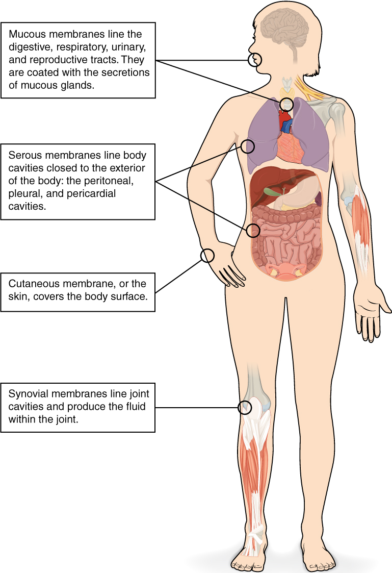
The connective tissue membrane    is formed solely from connective tissue. These membranes encapsulate organs, such as the kidneys, and line our movable joints. A synovial membrane    is a type of connective tissue membrane that lines the cavity of a freely movable joint. For example, synovial membranes surround the joints of the shoulder, elbow, and knee. Fibroblasts in the inner layer of the synovial membrane release hyaluronan into the joint cavity. The hyaluronan effectively traps available water to form the synovial fluid, a natural lubricant that enables the bones of a joint to move freely against one another without much friction. This synovial fluid readily exchanges water and nutrients with blood, as do all body fluids.

Epithelial membranes

The epithelial membrane    is composed of epithelium attached to a layer of connective tissue, for example, your skin. The mucous membrane    is also a composite of connective and epithelial tissues. Sometimes called mucosae, these epithelial membranes line the body cavities and hollow passageways that open to the external environment, and include the digestive, respiratory, excretory, and reproductive tracts. Mucous, produced by the epithelial exocrine glands, covers the epithelial layer. The underlying connective tissue, called the lamina propria    (literally “own layer”), help support the fragile epithelial layer.

A serous membrane    is an epithelial membrane composed of mesodermally derived epithelium called the mesothelium that is supported by connective tissue. These membranes line the coelomic cavities of the body, that is, those cavities that do not open to the outside, and they cover the organs located within those cavities. They are essentially membranous bags, with mesothelium lining the inside and connective tissue on the outside. Serous fluid secreted by the cells of the thin squamous mesothelium lubricates the membrane and reduces abrasion and friction between organs. Serous membranes are identified according locations. Three serous membranes line the thoracic cavity; the two pleura that cover the lungs and the pericardium that covers the heart. A fourth, the peritoneum, is the serous membrane in the abdominal cavity that covers abdominal organs and forms double sheets of mesenteries that suspend many of the digestive organs.

The skin is an epithelial membrane also called the cutaneous membrane    . It is a stratified squamous epithelial membrane resting on top of connective tissue. The apical surface of this membrane is exposed to the external environment and is covered with dead, keratinized cells that help protect the body from desiccation and pathogens.

Chapter review

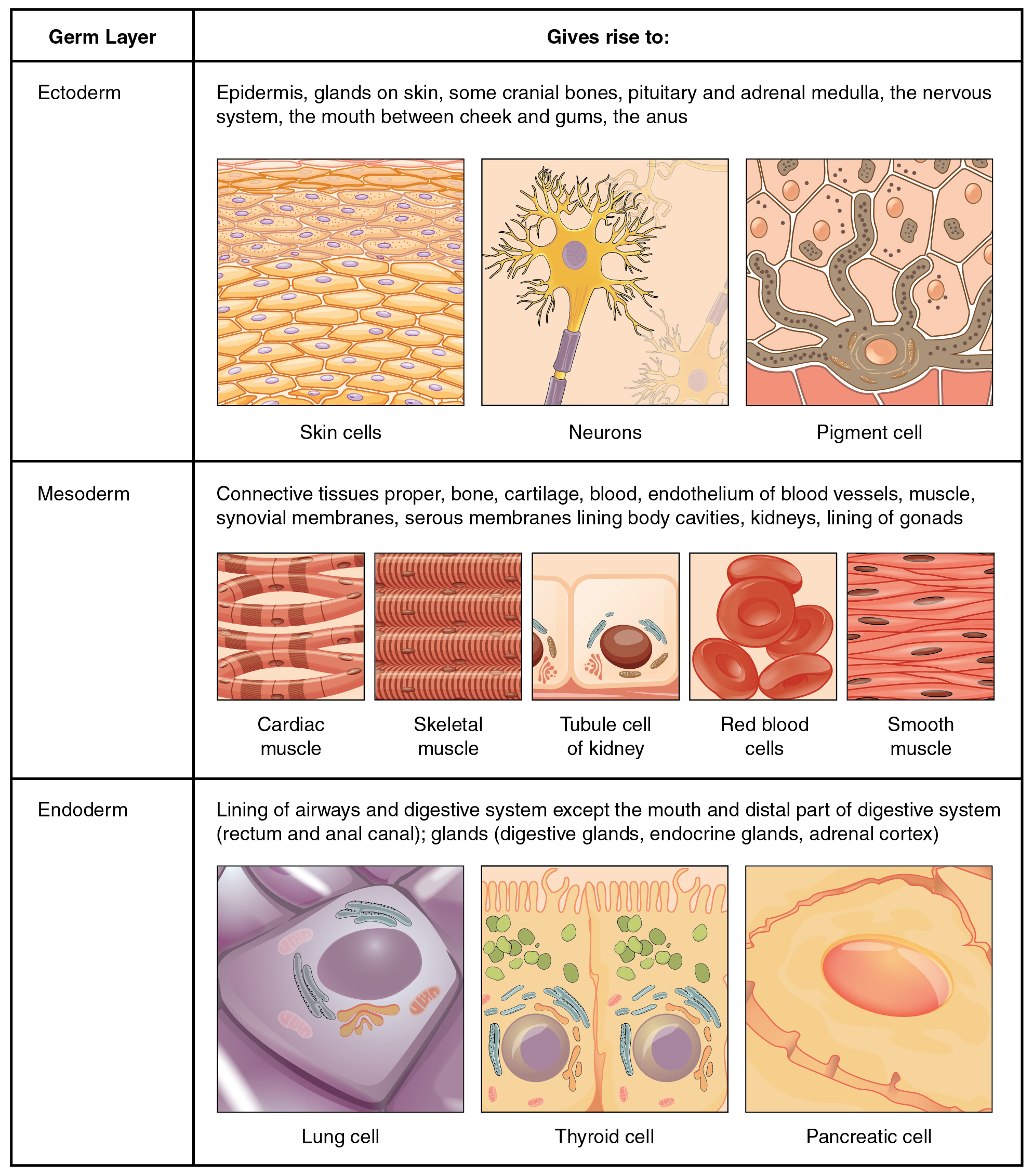
The human body contains more than 200 types of cells that can all be classified into four types of tissues: epithelial, connective, muscle, and nervous. Epithelial tissues act as coverings controlling the movement of materials across the surface. Connective tissue integrates the various parts of the body and provides support and protection to organs. Muscle tissue allows the body to move. Nervous tissues propagate information.

The study of the shape and arrangement of cells in tissue is called histology. All cells and tissues in the body derive from three germ layers in the embryo: the ectoderm, mesoderm, and endoderm.

Different types of tissues form membranes that enclose organs, provide a friction-free interaction between organs, and keep organs together. Synovial membranes are connective tissue membranes that protect and line the joints. Epithelial membranes are formed from epithelial tissue attached to a layer of connective tissue. There are three types of epithelial membranes: mucous, which contain glands; serous, which secrete fluid; and cutaneous which makes up the skin.

View this slideshow to learn more about stem cells. How do somatic stem cells differ from embryonic stem cells?

Most somatic stem cells give rise to only a few cell types.

Got questions? Get instant answers now!

Questions & Answers

A golfer on a fairway is 70 m away from the green, which sits below the level of the fairway by 20 m. If the golfer hits the ball at an angle of 40° with an initial speed of 20 m/s, how close to the green does she come?
Aislinn Reply
cm
tijani
what is titration
John Reply
what is physics
Siyaka Reply
A mouse of mass 200 g falls 100 m down a vertical mine shaft and lands at the bottom with a speed of 8.0 m/s. During its fall, how much work is done on the mouse by air resistance
Jude Reply
Can you compute that for me. Ty
Jude
what is the dimension formula of energy?
David Reply
what is viscosity?
David
what is inorganic
emma Reply
what is chemistry
Youesf Reply
what is inorganic
emma
Chemistry is a branch of science that deals with the study of matter,it composition,it structure and the changes it undergoes
Adjei
please, I'm a physics student and I need help in physics
Adjanou
chemistry could also be understood like the sexual attraction/repulsion of the male and female elements. the reaction varies depending on the energy differences of each given gender. + masculine -female.
Pedro
A ball is thrown straight up.it passes a 2.0m high window 7.50 m off the ground on it path up and takes 1.30 s to go past the window.what was the ball initial velocity
Krampah Reply
2. A sled plus passenger with total mass 50 kg is pulled 20 m across the snow (0.20) at constant velocity by a force directed 25° above the horizontal. Calculate (a) the work of the applied force, (b) the work of friction, and (c) the total work.
Sahid Reply
you have been hired as an espert witness in a court case involving an automobile accident. the accident involved car A of mass 1500kg which crashed into stationary car B of mass 1100kg. the driver of car A applied his brakes 15 m before he skidded and crashed into car B. after the collision, car A s
Samuel Reply
can someone explain to me, an ignorant high school student, why the trend of the graph doesn't follow the fact that the higher frequency a sound wave is, the more power it is, hence, making me think the phons output would follow this general trend?
Joseph Reply
Nevermind i just realied that the graph is the phons output for a person with normal hearing and not just the phons output of the sound waves power, I should read the entire thing next time
Joseph
Follow up question, does anyone know where I can find a graph that accuretly depicts the actual relative "power" output of sound over its frequency instead of just humans hearing
Joseph
"Generation of electrical energy from sound energy | IEEE Conference Publication | IEEE Xplore" ***ieeexplore.ieee.org/document/7150687?reload=true
Ryan
what's motion
Maurice Reply
what are the types of wave
Maurice
answer
Magreth
progressive wave
Magreth
hello friend how are you
Muhammad Reply
fine, how about you?
Mohammed
hi
Mujahid
A string is 3.00 m long with a mass of 5.00 g. The string is held taut with a tension of 500.00 N applied to the string. A pulse is sent down the string. How long does it take the pulse to travel the 3.00 m of the string?
yasuo Reply
Who can show me the full solution in this problem?
Reofrir Reply
Got questions? Join the online conversation and get instant answers!
Jobilize.com Reply

Get Jobilize Job Search Mobile App in your pocket Now!

Get it on Google Play Download on the App Store Now


Source:  OpenStax, Anatomy & Physiology. OpenStax CNX. Feb 04, 2016 Download for free at http://legacy.cnx.org/content/col11496/1.8
Google Play and the Google Play logo are trademarks of Google Inc.

Notification Switch

Would you like to follow the 'Anatomy & Physiology' conversation and receive update notifications?

Ask