

| << Chapter < Page | Chapter >> Page > |
At birth, the brain case and orbits of the skull are disproportionally large compared to the bones of the jaws and lower face. This reflects the relative underdevelopment of the maxilla and mandible, which lack teeth, and the small sizes of the paranasal sinuses and nasal cavity. During early childhood, the mastoid process enlarges, the two halves of the mandible and frontal bone fuse together to form single bones, and the paranasal sinuses enlarge. The jaws also expand as the teeth begin to appear. These changes all contribute to the rapid growth and enlargement of the face during childhood.
Development of the vertebrae begins with the accumulation of mesenchyme cells from each sclerotome around the notochord. These cells differentiate into a hyaline cartilage model for each vertebra, which then grow and eventually ossify into bone through the process of endochondral ossification. As the developing vertebrae grow, the notochord largely disappears. However, small areas of notochord tissue persist between the adjacent vertebrae and this contributes to the formation of each intervertebral disc.
The ribs and sternum also develop from mesenchyme. The ribs initially develop as part of the cartilage model for each vertebra, but in the thorax region, the rib portion separates from the vertebra by the eighth week. The cartilage model of the rib then ossifies, except for the anterior portion, which remains as the costal cartilage. The sternum initially forms as paired hyaline cartilage models on either side of the anterior midline, beginning during the fifth week of development. The cartilage models of the ribs become attached to the lateral sides of the developing sternum. Eventually, the two halves of the cartilaginous sternum fuse together along the midline and then ossify into bone. The manubrium and body of the sternum are converted into bone first, with the xiphoid process remaining as cartilage until late in life.
View this video to review the two processes that give rise to the bones of the skull and body. What are the two mechanisms by which the bones of the body are formed and which bones are formed by each mechanism?
The early fusion of a suture in primary craniosynostosis prevents any additional enlargement of the cranial bones and skull along this line. Continued growth of the brain and skull is therefore diverted to other areas of the head, causing an abnormal enlargement of these regions. For example, the early disappearance of the anterior fontanelle and premature closure of the sagittal suture prevents growth across the top of the head. This is compensated by upward growth by the bones of the lateral skull, resulting in a long, narrow, wedge-shaped head. This condition, known as scaphocephaly, accounts for approximately 50 percent of craniosynostosis abnormalities. Although the skull is misshapen, the brain still has adequate room to grow and thus there is no accompanying abnormal neurological development.
In cases of complex craniosynostosis, several sutures close prematurely. The amount and degree of skull deformity is determined by the location and extent of the sutures involved. This results in more severe constraints on skull growth, which can alter or impede proper brain growth and development.
Cases of craniosynostosis are usually treated with surgery. A team of physicians will open the skull along the fused suture, which will then allow the skull bones to resume their growth in this area. In some cases, parts of the skull will be removed and replaced with an artificial plate. The earlier after birth that surgery is performed, the better the outcome. After treatment, most children continue to grow and develop normally and do not exhibit any neurological problems.
Formation of the axial skeleton begins during early embryonic development with the appearance of the rod-like notochord along the dorsal length of the early embryo. Repeating, paired blocks of tissue called somites then appear along either side of notochord. As the somites grow, they split into parts, one of which is called a sclerotome. This consists of mesenchyme, the embryonic tissue that will become the bones, cartilages, and connective tissues of the body.
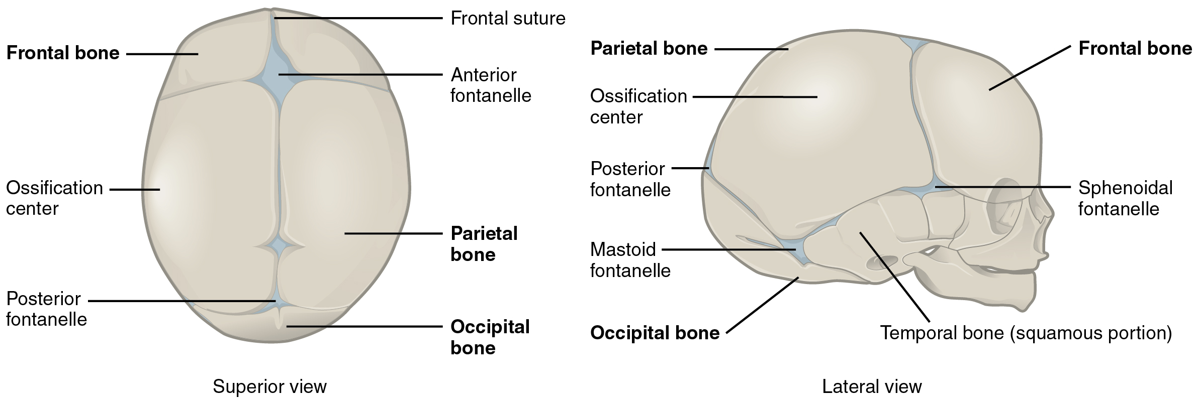
Mesenchyme in the head region will produce the bones of the skull via two different mechanisms. The bones of the brain case arise via intramembranous ossification in which embryonic mesenchyme tissue converts directly into bone. At the time of birth, these bones are separated by fontanelles, wide areas of fibrous connective tissue. As the bones grow, the fontanelles are reduced to sutures, which allow for continued growth of the skull throughout childhood. In contrast, the cranial base and facial bones are produced by the process of endochondral ossification, in which mesenchyme tissue initially produces a hyaline cartilage model of the future bone. The cartilage model allows for growth of the bone and is gradually converted into bone over a period of many years.
The vertebrae, ribs, and sternum also develop via endochondral ossification. Mesenchyme accumulates around the notochord and produces hyaline cartilage models of the vertebrae. The notochord largely disappears, but remnants of the notochord contribute to formation of the intervertebral discs. In the thorax region, a portion of the vertebral cartilage model splits off to form the ribs. These then become attached anteriorly to the developing cartilage model of the sternum. Growth of the cartilage models for the vertebrae, ribs, and sternum allow for enlargement of the thoracic cage during childhood and adolescence. The cartilage models gradually undergo ossification and are converted into bone.
View this video to review the two processes that give rise to the bones of the skull and body. What are the two mechanisms by which the bones of the body are formed and which bones are formed by each mechanism?
Bones on the top and sides of the skull develop when fibrous membrane areas ossify (convert) into bone. The bones of the limbs, ribs, and vertebrae develop when cartilage models of the bones ossify into bone.
Notification Switch
Would you like to follow the 'Anatomy & Physiology' conversation and receive update notifications?

|
|
|
|
|

|
|

|
|
|
|
|

|
|

|
|

|
|

|
|
|
|
|

|
|

|