<< Chapter < Page Chapter >> Page >

The lateral compartment of the leg    includes two muscles: the fibularis longus    (peroneus longus) and the fibularis brevis    (peroneus brevis). The superficial muscles in the posterior compartment of the leg    all insert onto the calcaneal tendon    (Achilles tendon), a strong tendon that inserts into the calcaneal bone of the ankle. The muscles in this compartment are large and strong and keep humans upright. The most superficial and visible muscle of the calf is the gastrocnemius    . Deep to the gastrocnemius is the wide, flat soleus    . The plantaris    runs obliquely between the two; some people may have two of these muscles, whereas no plantaris is observed in about seven percent of other cadaver dissections. The plantaris tendon is a desirable substitute for the fascia lata in hernia repair, tendon transplants, and repair of ligaments. There are four deep muscles in the posterior compartment of the leg as well: the popliteus    , flexor digitorum longus    , flexor hallucis longus    , and tibialis posterior    .

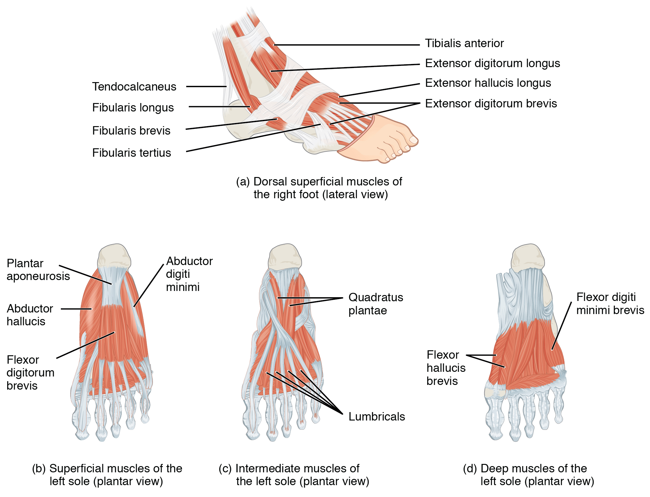
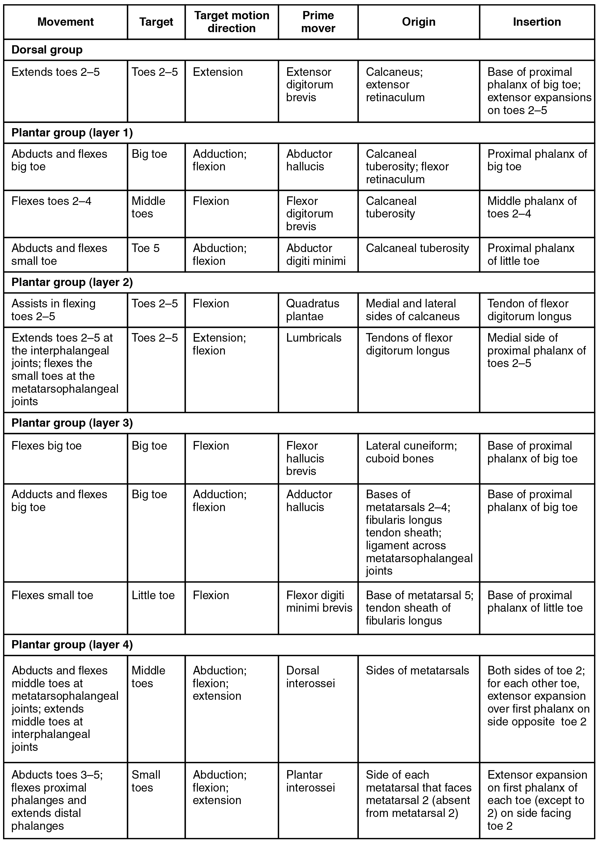
The foot also has intrinsic muscles, which originate and insert within it (similar to the intrinsic muscles of the hand). These muscles primarily provide support for the foot and its arch, and contribute to movements of the toes ( [link] and [link] ). The principal support for the longitudinal arch of the foot is a deep fascia called plantar aponeurosis    , which runs from the calcaneus bone to the toes (inflammation of this tissue is the cause of “plantar fasciitis,” which can affect runners. The intrinsic muscles of the foot consist of two groups. The dorsal group    includes only one muscle, the extensor digitorum brevis    . The second group is the plantar group    , which consists of four layers, starting with the most superficial.

Intrinsic muscles of the foot

This figure shows the muscles of the foot. The top panel shows the lateral view of the dorsal muscles. The bottom left panel shows the superficial muscles of the left sole, the center panel shows the intermediate muscles of the left sole, and the right panel shows the deep muscles of the left sole.
The muscles along the dorsal side of the foot (a) generally extend the toes while the muscles of the plantar side of the foot (b, c, d) generally flex the toes. The plantar muscles exist in three layers, providing the foot the strength to counterbalance the weight of the body. In this diagram, these three layers are shown from a plantar view beginning with the bottom-most layer just under the plantar skin of the foot (b) and ending with the top-most layer (d) located just inferior to the foot and toe bones.

Intrinsic muscles in the foot

This table describes intrinsic muscles in the foot. The dorsal group consists of the extensor digitorum brevis, which extends toes 2 through 5. It originates in the calcaneus and the extensor retinaculum. These muscles make up layer 1 of the plantar group. The abductor hallucis abducts and flexes the big toe. It originates in the calcaneal tuberosity and flexor retinaculum. The flexor digitorum brevis flexes toes 2 through 4. It originates in the calcaneal tuberosity. The abductor digiti minimi abducts and flexes the small toe. It originates in the calcaneal tuberosity. These muscles make up layer 2 of the plantar group. The quadratus plantae assists in flexing toes 2 through 5. It originates in the medial and lateral sides of the calcaneus. The lumbricals extend toes 2 through 5 at the interphalangeal joints; they also flex the small toes at the metatarsophalangeal joints. They originate in the tendons of the flexor digitorum longus. These muscles make up layer 3 of the plantar group. The flexor hallucis brevis flexes the big toe. It originates in the lateral cuneiform and in the cuboid bones. The adductor hallucis adducts and flexes the big toe. It originates in the bases of metatarsals 2 through 4, in the fibularis longus tendon sheath, and in the ligament across the metatarsophalangeal joints. The flexor digiti minimi brevis flexes the small toe. It originates in the base of metatarsal 5 and in the tendon sheath of the fibularis longus. These muscles make up layer 4 of the plantar group. The dorsal interossei abducts and flexes the middle toes at the metatarsophalangeal joints; it also extends the middle toes at the interphalangeal joints. It originates in the sides of the metatarsals. The plantar interossei abducts toes 3 through 5; it also flexes the proximal phalanges and extends the distal phalanges. It originates in the side of each metatarsal that faces metatarsal 2 (absent from metatarsal 2).

Chapter review

The pelvic girdle attaches the legs to the axial skeleton. The hip joint is where the pelvic girdle and the leg come together. The hip is joined to the pelvic girdle by many muscles. In the gluteal region, the psoas major and iliacus form the iliopsoas. The large and strong gluteus maximus, gluteus medius, and gluteus minimus extend and abduct the femur. Along with the gluteus maximus, the tensor fascia lata muscle forms the iliotibial tract. The lateral rotators of the femur at the hip are the piriformis, obturator internus, obturator externus, superior gemellus, inferior gemellus, and quadratus femoris. On the medial part of the thigh, the adductor longus, adductor brevis, and adductor magnus adduct the thigh and medially rotate it. The pectineus muscle adducts and flexes the femur at the hip.

The thigh muscles that move the femur, tibia, and fibula are divided into medial, anterior, and posterior compartments. The medial compartment includes the adductors, pectineus, and the gracilis. The anterior compartment comprises the quadriceps femoris, quadriceps tendon, patellar ligament, and the sartorius. The quadriceps femoris is made of four muscles: the rectus femoris, the vastus lateralis, the vastus medius, and the vastus intermedius, which together extend the knee. The posterior compartment of the thigh includes the hamstrings: the biceps femoris, semitendinosus, and the semimembranosus, which all flex the knee.

The muscles of the leg that move the foot and toes are divided into anterior, lateral, superficial- and deep-posterior compartments. The anterior compartment includes the tibialis anterior, the extensor hallucis longus, the extensor digitorum longus, and the fibularis (peroneus) tertius. The lateral compartment houses the fibularis (peroneus) longus and the fibularis (peroneus) brevis. The superficial posterior compartment has the gastrocnemius, soleus, and plantaris; and the deep posterior compartment has the popliteus, tibialis posterior, flexor digitorum longus, and flexor hallucis longus.

Questions & Answers

how does Neisseria cause meningitis
Nyibol Reply
what is microbiologist
Muhammad Reply
what is errata
Muhammad
is the branch of biology that deals with the study of microorganisms.
Ntefuni Reply
What is microbiology
Mercy Reply
studies of microbes
Louisiaste
when we takee the specimen which lumbar,spin,
Ziyad Reply
How bacteria create energy to survive?
Muhamad Reply
Bacteria doesn't produce energy they are dependent upon their substrate in case of lack of nutrients they are able to make spores which helps them to sustain in harsh environments
_Adnan
But not all bacteria make spores, l mean Eukaryotic cells have Mitochondria which acts as powerhouse for them, since bacteria don't have it, what is the substitution for it?
Muhamad
they make spores
Louisiaste
what is sporadic nd endemic, epidemic
Aminu Reply
the significance of food webs for disease transmission
Abreham
food webs brings about an infection as an individual depends on number of diseased foods or carriers dully.
Mark
explain assimilatory nitrate reduction
Esinniobiwa Reply
Assimilatory nitrate reduction is a process that occurs in some microorganisms, such as bacteria and archaea, in which nitrate (NO3-) is reduced to nitrite (NO2-), and then further reduced to ammonia (NH3).
Elkana
This process is called assimilatory nitrate reduction because the nitrogen that is produced is incorporated in the cells of microorganisms where it can be used in the synthesis of amino acids and other nitrogen products
Elkana
Examples of thermophilic organisms
Shu Reply
Give Examples of thermophilic organisms
Shu
advantages of normal Flora to the host
Micheal Reply
Prevent foreign microbes to the host
Abubakar
they provide healthier benefits to their hosts
ayesha
They are friends to host only when Host immune system is strong and become enemies when the host immune system is weakened . very bad relationship!
Mark
what is cell
faisal Reply
cell is the smallest unit of life
Fauziya
cell is the smallest unit of life
Akanni
ok
Innocent
cell is the structural and functional unit of life
Hasan
is the fundamental units of Life
Musa
what are emergency diseases
Micheal Reply
There are nothing like emergency disease but there are some common medical emergency which can occur simultaneously like Bleeding,heart attack,Breathing difficulties,severe pain heart stock.Hope you will get my point .Have a nice day ❣️
_Adnan
define infection ,prevention and control
Innocent
I think infection prevention and control is the avoidance of all things we do that gives out break of infections and promotion of health practices that promote life
Lubega
Heyy Lubega hussein where are u from?
_Adnan
en français
Adama
which site have a normal flora
ESTHER Reply
Many sites of the body have it Skin Nasal cavity Oral cavity Gastro intestinal tract
Safaa
skin
Asiina
skin,Oral,Nasal,GIt
Sadik
How can Commensal can Bacteria change into pathogen?
Sadik
How can Commensal Bacteria change into pathogen?
Sadik
all
Tesfaye
by fussion
Asiina
what are the advantages of normal Flora to the host
Micheal
what are the ways of control and prevention of nosocomial infection in the hospital
Micheal
what is inflammation
Shelly Reply
part of a tissue or an organ being wounded or bruised.
Wilfred
what term is used to name and classify microorganisms?
Micheal Reply
Binomial nomenclature
adeolu
Got questions? Join the online conversation and get instant answers!
Jobilize.com Reply

Get Jobilize Job Search Mobile App in your pocket Now!

Get it on Google Play Download on the App Store Now


Source:  OpenStax, Anatomy & Physiology. OpenStax CNX. Feb 04, 2016 Download for free at http://legacy.cnx.org/content/col11496/1.8
Google Play and the Google Play logo are trademarks of Google Inc.

Notification Switch

Would you like to follow the 'Anatomy & Physiology' conversation and receive update notifications?

Ask