<< Chapter < Page Chapter >> Page >
  • The eight incisors , four top and four bottom, are the sharp front teeth you use for biting into food.
  • The four cuspids (or canines) flank the incisors and have a pointed edge (cusp) to tear up food. These fang-like teeth are superb for piercing tough or fleshy foods.
  • Posterior to the cuspids are the eight premolars (or bicuspids), which have an overall flatter shape with two rounded cusps useful for mashing foods.
  • The most posterior and largest are the 12 molars , which have several pointed cusps used to crush food so it is ready for swallowing. The third members of each set of three molars, top and bottom, are commonly referred to as the wisdom teeth, because their eruption is commonly delayed until early adulthood. It is not uncommon for wisdom teeth to fail to erupt; that is, they remain impacted. In these cases, the teeth are typically removed by orthodontic surgery.

Permanent and deciduous teeth

This diagram shows the arrangement of permanent and deciduous teeth in human. The permanent teeth are labeled along with the average age at which they emerge. An inset shows the arrangement of the deciduous teeth, with the age at which they emerge listed.
This figure of two human dentitions shows the arrangement of teeth in the maxilla and mandible, and the relationship between the deciduous and permanent teeth.

Anatomy of a tooth

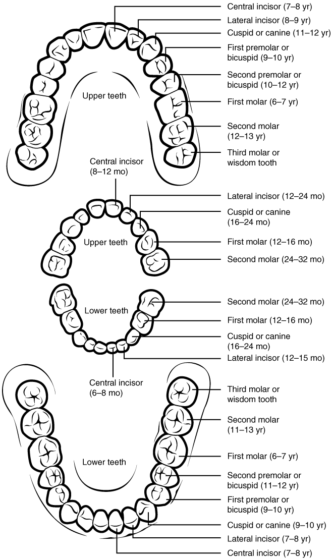
The teeth are secured in the alveolar processes (sockets) of the maxilla and the mandible. Gingivae (commonly called the gums) are soft tissues that line the alveolar processes and surround the necks of the teeth. Teeth are also held in their sockets by a connective tissue called the periodontal ligament.

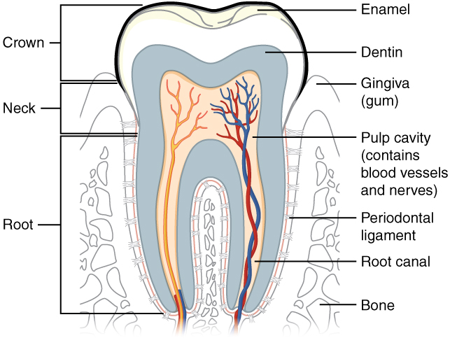
The two main parts of a tooth are the crown    , which is the portion projecting above the gum line, and the root    , which is embedded within the maxilla and mandible. Both parts contain an inner pulp cavity    , containing loose connective tissue through which run nerves and blood vessels. The region of the pulp cavity that runs through the root of the tooth is called the root canal. Surrounding the pulp cavity is dentin    , a bone-like tissue. In the root of each tooth, the dentin is covered by an even harder bone-like layer called cementum    . In the crown of each tooth, the dentin is covered by an outer layer of enamel    , the hardest substance in the body ( [link] ).

Although enamel protects the underlying dentin and pulp cavity, it is still nonetheless susceptible to mechanical and chemical erosion, or what is known as tooth decay. The most common form, dental caries (cavities) develops when colonies of bacteria feeding on sugars in the mouth release acids that cause soft tissue inflammation and degradation of the calcium crystals of the enamel. The digestive functions of the mouth are summarized in [link] .

The structure of the tooth

This diagram shows a cross-section of a human tooth elucidating its structure. The major parts of the tooth along with the blood vessels are shown.
This longitudinal section through a molar in its alveolar socket shows the relationships between enamel, dentin, and pulp.
Digestive Functions of the Mouth
Structure Action Outcome
Lips and cheeks Confine food between teeth
  • Food is chewed evenly during mastication
Salivary glands Secrete saliva
  • Moisten and lubricate the lining of the mouth and pharynx
  • Moisten, soften, and dissolve food
  • Clean the mouth and teeth
  • Salivary amylase breaks down starch
Tongue’s extrinsic muscles Move tongue sideways, and in and out
  • Manipulate food for chewing
  • Shape food into a bolus
  • Manipulate food for swallowing
Tongue’s intrinsic muscles Change tongue shape
  • Manipulate food for swallowing
Taste buds Sense food in mouth and sense taste
  • Nerve impulses from taste buds are conducted to salivary nuclei in the brain stem and then to salivary glands, stimulating saliva secretion
Lingual glands Secrete lingual lipase
  • Activated in the stomach
  • Break down triglycerides into fatty acids and diglycerides
Teeth Shred and crush food
  • Break down solid food into smaller particles for deglutition

Questions & Answers

what is phylogeny
Odigie Reply
evolutionary history and relationship of an organism or group of organisms
AI-Robot
ok
Deng
what is biology
Hajah Reply
the study of living organisms and their interactions with one another and their environments
AI-Robot
what is biology
Victoria Reply
HOW CAN MAN ORGAN FUNCTION
Alfred Reply
the diagram of the digestive system
Assiatu Reply
allimentary cannel
Ogenrwot
How does twins formed
William Reply
They formed in two ways first when one sperm and one egg are splited by mitosis or two sperm and two eggs join together
Oluwatobi
what is genetics
Josephine Reply
Genetics is the study of heredity
Misack
how does twins formed?
Misack
What is manual
Hassan Reply
discuss biological phenomenon and provide pieces of evidence to show that it was responsible for the formation of eukaryotic organelles
Joseph Reply
what is biology
Yousuf Reply
the study of living organisms and their interactions with one another and their environment.
Wine
discuss the biological phenomenon and provide pieces of evidence to show that it was responsible for the formation of eukaryotic organelles in an essay form
Joseph Reply
what is the blood cells
Shaker Reply
list any five characteristics of the blood cells
Shaker
lack electricity and its more savely than electronic microscope because its naturally by using of light
Abdullahi Reply
advantage of electronic microscope is easily and clearly while disadvantage is dangerous because its electronic. advantage of light microscope is savely and naturally by sun while disadvantage is not easily,means its not sharp and not clear
Abdullahi
cell theory state that every organisms composed of one or more cell,cell is the basic unit of life
Abdullahi
is like gone fail us
DENG
cells is the basic structure and functions of all living things
Ramadan
What is classification
ISCONT Reply
is organisms that are similar into groups called tara
Yamosa
in what situation (s) would be the use of a scanning electron microscope be ideal and why?
Kenna Reply
A scanning electron microscope (SEM) is ideal for situations requiring high-resolution imaging of surfaces. It is commonly used in materials science, biology, and geology to examine the topography and composition of samples at a nanoscale level. SEM is particularly useful for studying fine details,
Hilary
Got questions? Join the online conversation and get instant answers!
Jobilize.com Reply

Get Jobilize Job Search Mobile App in your pocket Now!

Get it on Google Play Download on the App Store Now


Source:  OpenStax, Anatomy & Physiology. OpenStax CNX. Feb 04, 2016 Download for free at http://legacy.cnx.org/content/col11496/1.8
Google Play and the Google Play logo are trademarks of Google Inc.

Notification Switch

Would you like to follow the 'Anatomy & Physiology' conversation and receive update notifications?

Ask