<< Chapter < Page Chapter >> Page >
By the end of this section, you will be able to:
  • Describe the development of blood vessels
  • Describe the fetal circulation

In a developing embryo,the heart has developed enough by day 21 post-fertilization to begin beating. Circulation patterns are clearly established by the fourth week of embryonic life. It is critical to the survival of the developing human that the circulatory system forms early to supply the growing tissue with nutrients and gases, and to remove waste products. Blood cells and vessel production in structures outside the embryo proper called the yolk sac, chorion, and connecting stalk begin about 15 to 16 days following fertilization. Development of these circulatory elements within the embryo itself begins approximately 2 days later. You will learn more about the formation and function of these early structures when you study the chapter on development. During those first few weeks, blood vessels begin to form from the embryonic mesoderm. The precursor cells are known as hemangioblasts    . These in turn differentiate into angioblasts    , which give rise to the blood vessels and pluripotent stem cells, which differentiate into the formed elements of blood. (Seek additional content for more detail on fetal development and circulation.) Together, these cells form masses known as blood islands    scattered throughout the embryonic disc. Spaces appear on the blood islands that develop into vessel lumens. The endothelial lining of the vessels arise from the angioblasts within these islands. Surrounding mesenchymal cells give rise to the smooth muscle and connective tissue layers of the vessels. While the vessels are developing, the pluripotent stem cells begin to form the blood.

Vascular tubes also develop on the blood islands, and they eventually connect to one another as well as to the developing, tubular heart. Thus, the developmental pattern, rather than beginning from the formation of one central vessel and spreading outward, occurs in many regions simultaneously with vessels later joining together. This angiogenesis    —the creation of new blood vessels from existing ones—continues as needed throughout life as we grow and develop.

Blood vessel development often follows the same pattern as nerve development and travels to the same target tissues and organs. This occurs because the many factors directing growth of nerves also stimulate blood vessels to follow a similar pattern. Whether a given vessel develops into an artery or a vein is dependent upon local concentrations of signaling proteins.

As the embryo grows within the mother’s uterus, its requirements for nutrients and gas exchange also grow. The placenta—a circulatory organ unique to pregnancy—develops jointly from the embryo and uterine wall structures to fill this need. Emerging from the placenta is the umbilical vein    , which carries oxygen-rich blood from the mother to the fetal inferior vena cava via the ductus venosus to the heart that pumps it into fetal circulation. Two umbilical arteries    carry oxygen-depleted fetal blood, including wastes and carbon dioxide, to the placenta. Remnants of the umbilical arteries remain in the adult. (Seek additional content for more information on the role of the placenta in fetal circulation.)

There are three major shunts—alternate paths for blood flow—found in the circulatory system of the fetus. Two of these shunts divert blood from the pulmonary to the systemic circuit, whereas the third connects the umbilical vein to the inferior vena cava. The first two shunts are critical during fetal life, when the lungs are compressed, filled with amniotic fluid, and nonfunctional, and gas exchange is provided by the placenta. These shunts close shortly after birth, however, when the newborn begins to breathe. The third shunt persists a bit longer but becomes nonfunctional once the umbilical cord is severed. The three shunts are as follows ( [link] ):

  • The foramen ovale    is an opening in the interatrial septum that allows blood to flow from the right atrium to the left atrium. A valve associated with this opening prevents backflow of blood during the fetal period. As the newborn begins to breathe and blood pressure in the atria increases, this shunt closes. The fossa ovalis remains in the interatrial septum after birth, marking the location of the former foramen ovale.
  • The ductus arteriosus    is a short, muscular vessel that connects the pulmonary trunk to the aorta. Most of the blood pumped from the right ventricle into the pulmonary trunk is thereby diverted into the aorta. Only enough blood reaches the fetal lungs to maintain the developing lung tissue. When the newborn takes the first breath, pressure within the lungs drops dramatically, and both the lungs and the pulmonary vessels expand. As the amount of oxygen increases, the smooth muscles in the wall of the ductus arteriosus constrict, sealing off the passage. Eventually, the muscular and endothelial components of the ductus arteriosus degenerate, leaving only the connective tissue component of the ligamentum arteriosum.
  • The ductus venosus    is a temporary blood vessel that branches from the umbilical vein, allowing much of the freshly oxygenated blood from the placenta—the organ of gas exchange between the mother and fetus—to bypass the fetal liver and go directly to the fetal heart. The ductus venosus closes slowly during the first weeks of infancy and degenerates to become the ligamentum venosum.

Fetal shunts

This figure shows the blood vessels in a fetus.
The foramen ovale in the interatrial septum allows blood to flow from the right atrium to the left atrium. The ductus arteriosus is a temporary vessel, connecting the aorta to the pulmonary trunk. The ductus venosus links the umbilical vein to the inferior vena cava largely through the liver.

Chapter review

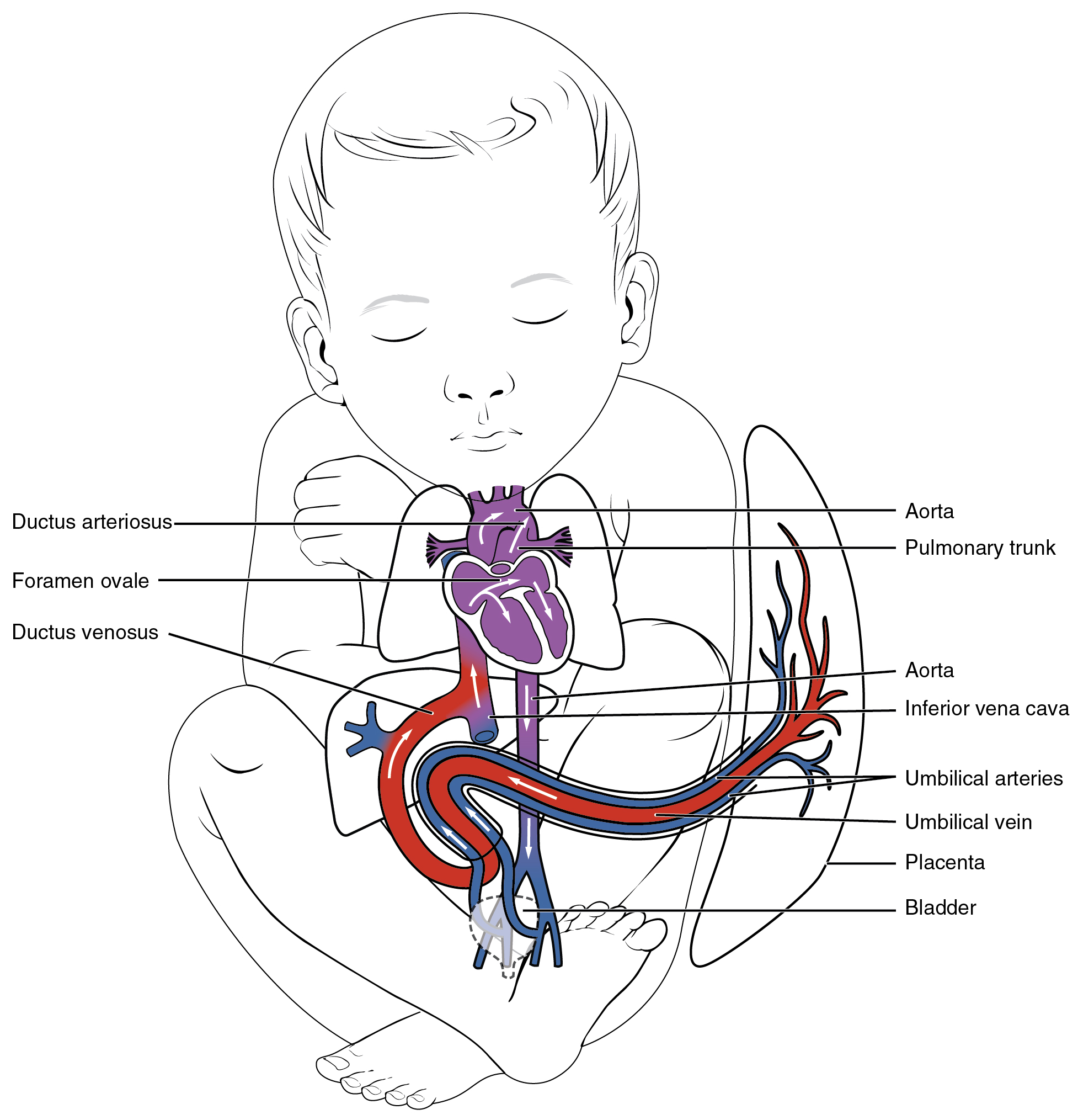
Blood vessels begin to form from the embryonic mesoderm. The precursor hemangioblasts differentiate into angioblasts, which give rise to the blood vessels and pluripotent stem cells that differentiate into the formed elements of the blood. Together, these cells form blood islands scattered throughout the embryo. Extensions known as vascular tubes eventually connect the vascular network. As the embryo grows within the mother’s womb, the placenta develops to supply blood rich in oxygen and nutrients via the umbilical vein and to remove wastes in oxygen-depleted blood via the umbilical arteries. Three major shunts found in the fetus are the foramen ovale and ductus arteriosus, which divert blood from the pulmonary to the systemic circuit, and the ductus venosus, which carries freshly oxygenated blood high in nutrients to the fetal heart.

Questions & Answers

how does Neisseria cause meningitis
Nyibol Reply
what is microbiologist
Muhammad Reply
what is errata
Muhammad
is the branch of biology that deals with the study of microorganisms.
Ntefuni Reply
What is microbiology
Mercy Reply
studies of microbes
Louisiaste
when we takee the specimen which lumbar,spin,
Ziyad Reply
How bacteria create energy to survive?
Muhamad Reply
Bacteria doesn't produce energy they are dependent upon their substrate in case of lack of nutrients they are able to make spores which helps them to sustain in harsh environments
_Adnan
But not all bacteria make spores, l mean Eukaryotic cells have Mitochondria which acts as powerhouse for them, since bacteria don't have it, what is the substitution for it?
Muhamad
they make spores
Louisiaste
what is sporadic nd endemic, epidemic
Aminu Reply
the significance of food webs for disease transmission
Abreham
food webs brings about an infection as an individual depends on number of diseased foods or carriers dully.
Mark
explain assimilatory nitrate reduction
Esinniobiwa Reply
Assimilatory nitrate reduction is a process that occurs in some microorganisms, such as bacteria and archaea, in which nitrate (NO3-) is reduced to nitrite (NO2-), and then further reduced to ammonia (NH3).
Elkana
This process is called assimilatory nitrate reduction because the nitrogen that is produced is incorporated in the cells of microorganisms where it can be used in the synthesis of amino acids and other nitrogen products
Elkana
Examples of thermophilic organisms
Shu Reply
Give Examples of thermophilic organisms
Shu
advantages of normal Flora to the host
Micheal Reply
Prevent foreign microbes to the host
Abubakar
they provide healthier benefits to their hosts
ayesha
They are friends to host only when Host immune system is strong and become enemies when the host immune system is weakened . very bad relationship!
Mark
what is cell
faisal Reply
cell is the smallest unit of life
Fauziya
cell is the smallest unit of life
Akanni
ok
Innocent
cell is the structural and functional unit of life
Hasan
is the fundamental units of Life
Musa
what are emergency diseases
Micheal Reply
There are nothing like emergency disease but there are some common medical emergency which can occur simultaneously like Bleeding,heart attack,Breathing difficulties,severe pain heart stock.Hope you will get my point .Have a nice day ❣️
_Adnan
define infection ,prevention and control
Innocent
I think infection prevention and control is the avoidance of all things we do that gives out break of infections and promotion of health practices that promote life
Lubega
Heyy Lubega hussein where are u from?
_Adnan
en français
Adama
which site have a normal flora
ESTHER Reply
Many sites of the body have it Skin Nasal cavity Oral cavity Gastro intestinal tract
Safaa
skin
Asiina
skin,Oral,Nasal,GIt
Sadik
How can Commensal can Bacteria change into pathogen?
Sadik
How can Commensal Bacteria change into pathogen?
Sadik
all
Tesfaye
by fussion
Asiina
what are the advantages of normal Flora to the host
Micheal
what are the ways of control and prevention of nosocomial infection in the hospital
Micheal
what is inflammation
Shelly Reply
part of a tissue or an organ being wounded or bruised.
Wilfred
what term is used to name and classify microorganisms?
Micheal Reply
Binomial nomenclature
adeolu
Got questions? Join the online conversation and get instant answers!
Jobilize.com Reply

Get Jobilize Job Search Mobile App in your pocket Now!

Get it on Google Play Download on the App Store Now


Source:  OpenStax, Anatomy & Physiology. OpenStax CNX. Feb 04, 2016 Download for free at http://legacy.cnx.org/content/col11496/1.8
Google Play and the Google Play logo are trademarks of Google Inc.

Notification Switch

Would you like to follow the 'Anatomy & Physiology' conversation and receive update notifications?

Ask