<< Chapter < Page Chapter >> Page >

Ion channels can also be specified by the diameter of the pore. The distance between the amino acids will be specific for the diameter of the ion when it dissociates from the water molecules surrounding it. Because of the surrounding water molecules, larger pores are not ideal for smaller ions because the water molecules will interact, by hydrogen bonds, more readily than the amino acid side chains. This is called size exclusion    . Some ion channels are selective for charge but not necessarily for size, and thus are called a nonspecific channel    . These nonspecific channels allow cations—particularly Na + , K + , and Ca 2+ —to cross the membrane, but exclude anions.

Ion channels do not always freely allow ions to diffuse across the membrane. Some are opened by certain events, meaning the channels are gated    . So another way that channels can be categorized is on the basis of how they are gated. Although these classes of ion channels are found primarily in the cells of nervous or muscular tissue, they also can be found in the cells of epithelial and connective tissues.

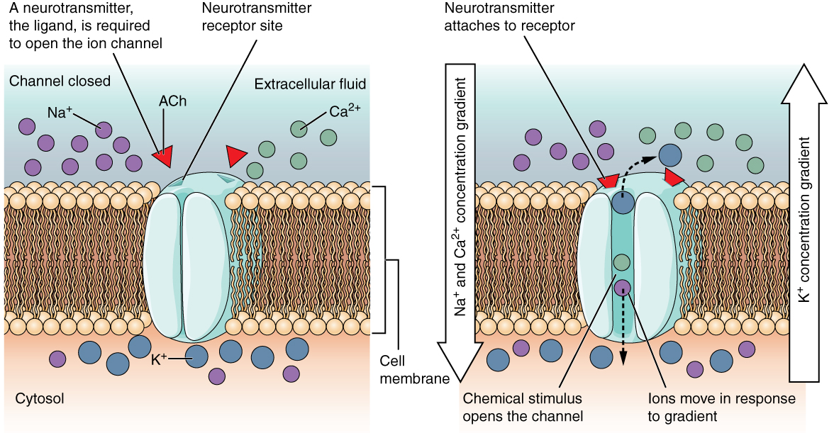
A ligand-gated channel opens because a signaling molecule, a ligand, binds to the extracellular region of the channel. This type of channel is also known as an ionotropic receptor    because when the ligand, known as a neurotransmitter in the nervous system, binds to the protein, ions cross the membrane changing its charge ( [link] ).

Ligand-gated channels

These two diagrams each show a channel protein embedded in the cell membrane. In the left diagram, there is a large number of sodium ions (NA plus) and calcium ions (CA two plus) in the extracellular fluid. Within the cytosol, there is a large number of potassium ions (K plus) but only a few sodium ions. In this diagram, the channel is closed. Two ACH molecules are floating in the extracellular fluid. Their label indicates that a neurotransmitter, a ligand, is required to open the ion channel. The neurotransmitter receptor site on the extracellular fluid side of the channel protein matches the shape of the ACH molecules. In the right diagram, the two ACH molecules attach to the neurotransmitter receptor sites on the channel protein. This opens the channel and the sodium and calcium ions diffuse through the channel and into the cytosol, down their concentration gradient. The potassium ions also diffuse through the channel in the opposite direction down their concentration gradient (out of the cell and into the extracellular fluid).
When the ligand, in this case the neurotransmitter acetylcholine, binds to a specific location on the extracellular surface of the channel protein, the pore opens to allow select ions through. The ions, in this case, are cations of sodium, calcium, and potassium.

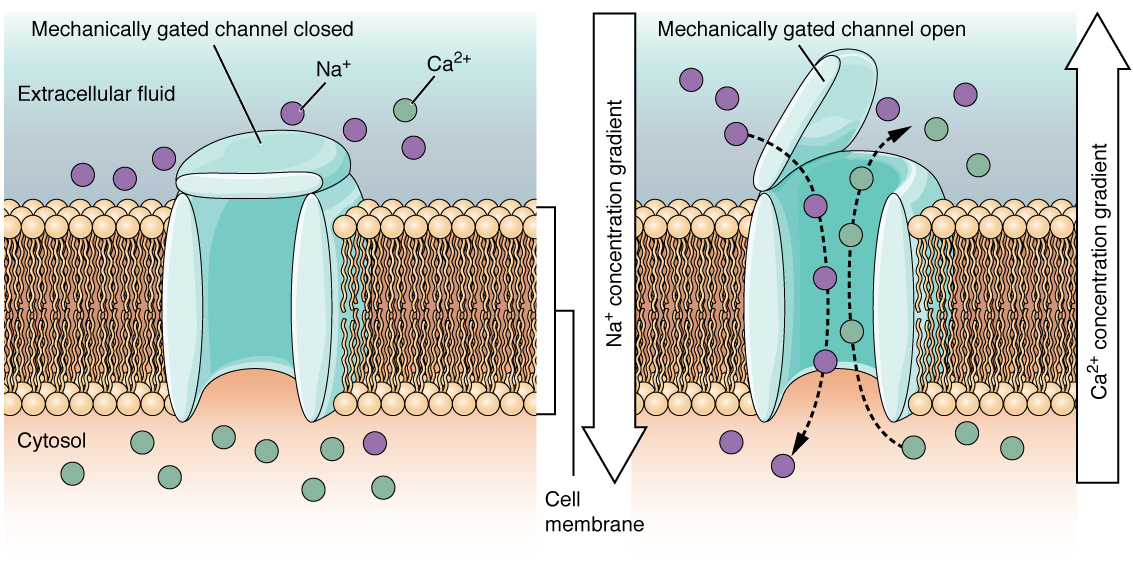
A mechanically gated channel    opens because of a physical distortion of the cell membrane. Many channels associated with the sense of touch (somatosensation) are mechanically gated. For example, as pressure is applied to the skin, these channels open and allow ions to enter the cell. Similar to this type of channel would be the channel that opens on the basis of temperature changes, as in testing the water in the shower ( [link] ).

Mechanically gated channels

These two diagrams each show a channel protein embedded in the cell membrane. In the left diagram, there are a large number of sodium ions in the extracellular fluid, but only a few sodium ions in the cytosol. There is a large number of calcium ions in the cytosol but only a few calcium ions in the extracellular fluid. In this diagram, the channel is closed, as the extracellular side has a lid, somewhat resembling that on a trash can, that is closed over the channel opening. In the right diagram, the mechanically gated channel is open.  This allows the sodium ions to flow from the extracellular fluid into the cell, down their concentration gradient. At the same time, the calcium ions are moving from the cytosol into the extracellular fluid, down their concentration gradient.
When a mechanical change occurs in the surrounding tissue, such as pressure or touch, the channel is physically opened. Thermoreceptors work on a similar principle. When the local tissue temperature changes, the protein reacts by physically opening the channel.

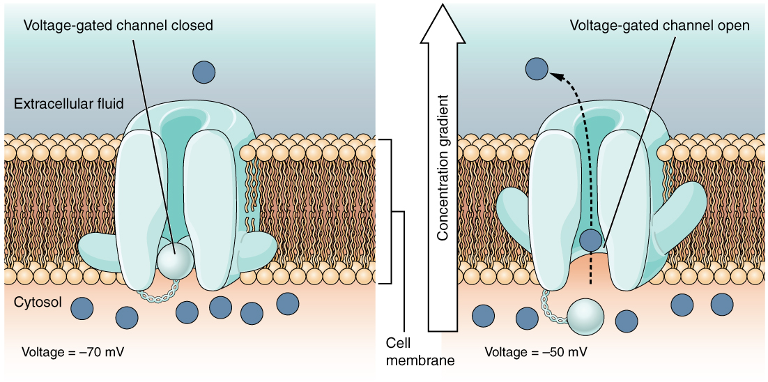
A voltage-gated channel    is a channel that responds to changes in the electrical properties of the membrane in which it is embedded. Normally, the inner portion of the membrane is at a negative voltage. When that voltage becomes less negative, the channel begins to allow ions to cross the membrane ( [link] ).

Voltage-gated channels

This is a two part diagram. Both diagrams show a voltage gated channel embedded in the lipid membrane bilayer. The channel contains a sphere shaped gate that is attached to a filament. In the first diagram there are several ions in the cytosol but only one ion in the extracellular fluid. The voltage across the membrane is currently minus seventy millivolts and the voltage gated channel is closed. In the second diagram, the voltage in the cytosol is minus fifty millivolts. This voltage change has caused the voltage gated channel to open, as the small sphere is no longer occluding the channel. One of the ions is moving through the channel, down its concentration gradient, and out into the extracellular fluid.
Voltage-gated channels open when the transmembrane voltage changes around them. Amino acids in the structure of the protein are sensitive to charge and cause the pore to open to the selected ion.

A leakage channel    is randomly gated, meaning that it opens and closes at random, hence the reference to leaking. There is no actual event that opens the channel; instead, it has an intrinsic rate of switching between the open and closed states. Leakage channels contribute to the resting transmembrane voltage of the excitable membrane ( [link] ).

Get Jobilize Job Search Mobile App in your pocket Now!

Get it on Google Play Download on the App Store Now


Source:  OpenStax, Anatomy & Physiology. OpenStax CNX. Feb 04, 2016 Download for free at http://legacy.cnx.org/content/col11496/1.8
Google Play and the Google Play logo are trademarks of Google Inc.

Notification Switch

Would you like to follow the 'Anatomy & Physiology' conversation and receive update notifications?

Ask